
|
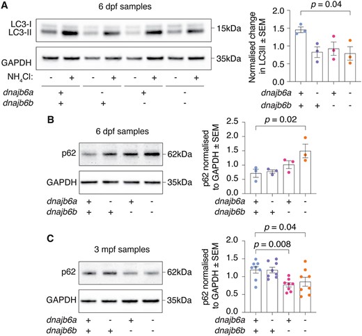
Fig. 4 Analysis of autophagy markers reveals early defect in double mutants. (A) Double mutant fish show a significant reduction in autophagic flux activity at 6 dpf based on difference in LC3-II levels between NH4Cl treated and untreated zebrafish. Experiment was completed in triplicate. (B) Western blot at 6 dpf shows a significant accumulation of p62 in double mutants. Error bars represent SEM for three biological replicates. (C) At 3 mpf dnajb6b single mutants and double mutants show a significant reduction in p62 levels compared to wildtype. Error bars represent SEM for eight fish across 3 biological replicates. GAPDH was used as a loading control and P62 values are normalised to GAPDH. Datasets were analysed with a one-way ANOVA and a two-way Dunnett multiple comparisons test.