
|
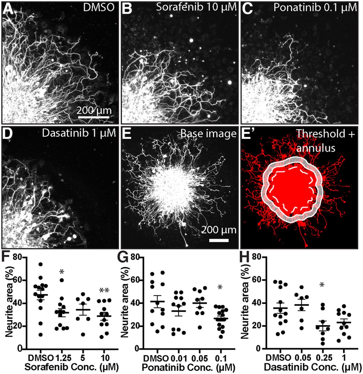
Fig. 8 MKI treatment induces loss of neurite density in mammalian DRGs. A–D, Single quadrant images of fixed DRG explants immunostained for βIII-Tubulin treated with vehicle or MKI compounds. MKI treatment led to shorter and less dense neurites. E, E', Demonstration of neurite area analysis. Immunostained DRG explants were imaged (E), thresholded (E', red), and then the explant core was identified (dotted white line) and a 50-μm-thick annulus (white shaded area) was created by drawing the inner and outer perimeter 50 and 100 μm from the edge of the explant core, respectively. F-H, Quantification of neurite area, analyzed by one-way ANOVA with post hoc Dunnett's test. F, Sorafenib: DMSO = 47.3 ± 4.2% area, 1.25 μm 32.0 ± 3.6, p = 0.016, 5 μm 34.5 ± 4.8, p = 0.116, 10 μm 28.8 ± 3.4, p = 0.0039. G, Ponatinib: DMSO = 41.4 ± 5.3, 0.01 μm 33.3 ± 4.4, p = 0.353, 0.05 μm 40.1 ± 3.9, p = 0.99, 1 μm 26.8 ± 2.2 p = 0.0215. H, Dasatinib: DMSO = 35.6 ± 4.5, 0.05 μm 38.4 ± 4.9, p = 0.954, 0.25 μm 20.0 ± 4.3, p = 0.038, 1 μm 23.0 ± 3.1, p = 0.073. Error bars represent SEM; *p < 0.05, **p < 0.01.