
|
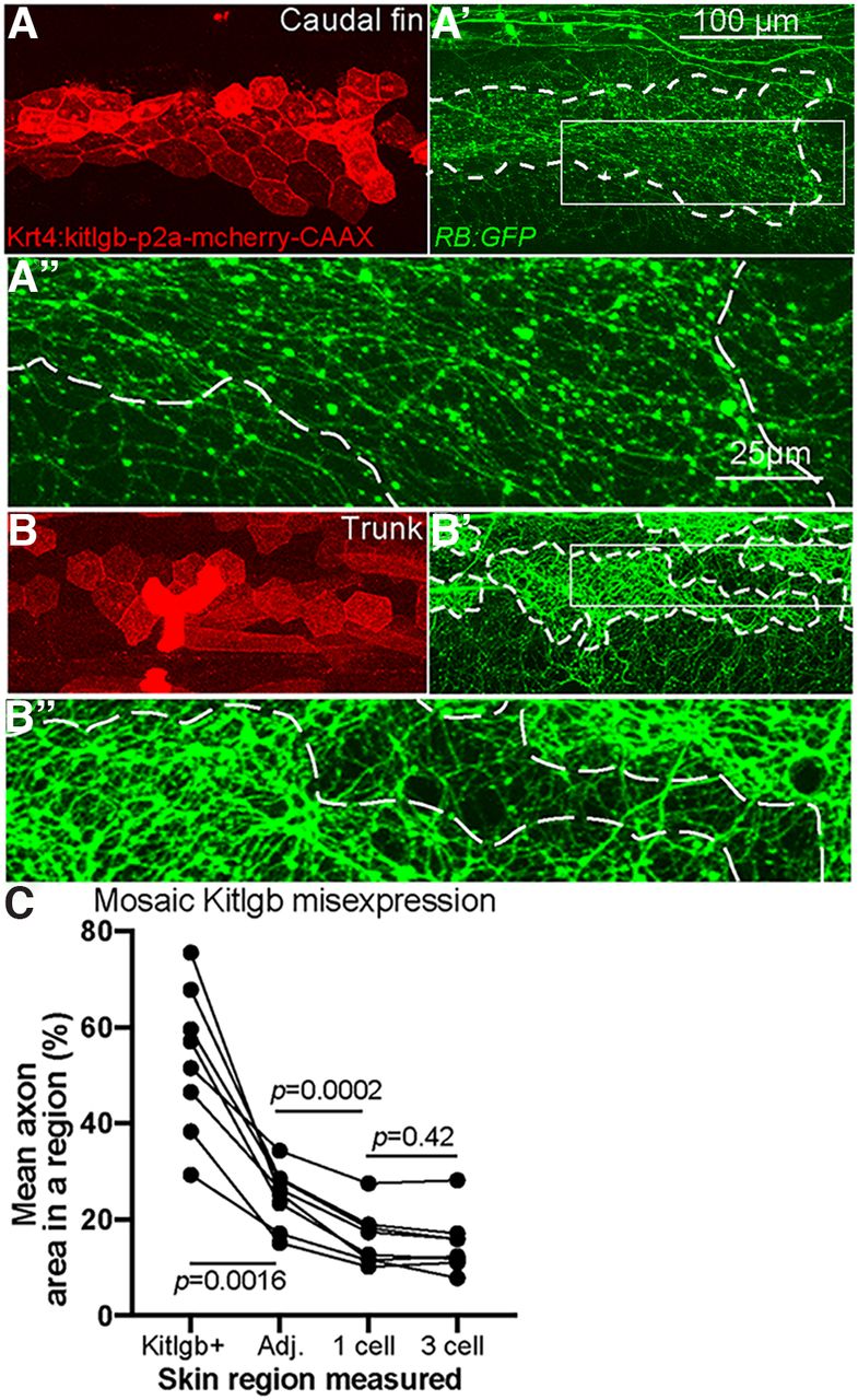
Fig. 5 Overexpression of Kit ligand b increases local axon density. A, B, Live imaging at 4 dpf of larvae injected with plasmid expressing kitlgb under a skin promoter. A', B', RB axons, labeled by RB:GFP, are significantly denser in regions of skin cells overexpressing kitlgb (white dashes indicate Kitlgb/mCherry+ regions). A”, B”, 4× magnified insets of A', B'. C, Quantification of axon density within individual larvae mosaically expressing Kitlgb (repeated measures ANOVA). Comparison of average axonal area within Kitlgb/mCherry+ regions (Kitlgb+), directly adjacent regions (Adj.), 1 cell length distance or 3 skin cell length distance from mCherry+ skin cells. Kitlgb+ = 53.2 ± 5.4% axonal area, Kitlgb–adjacent region =24.8 ± 2.2, 1 cell length distance = 16.0 ± 2.0, 3 cell length distance =15 ± 2.2. Kitlgb-misexpressing regions had significantly higher axonal density than any other region, while directly adjacent regions also had higher density than regions further away