
|
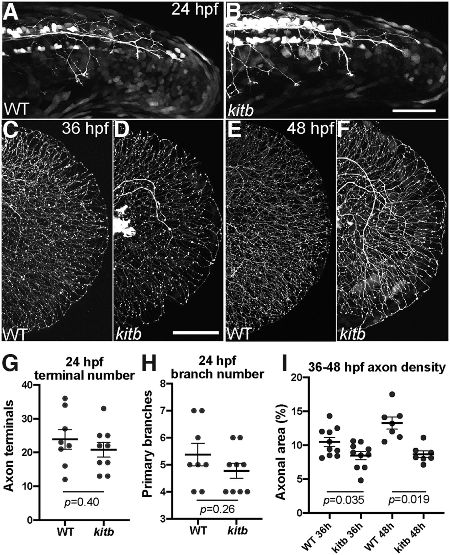
Fig. 4 Kitb mutants exhibit reduced axon density following normal initial extension. A–F, Live lateral images of RB:GFP tail regions from 24 to 48 hpf. Scale bar: 50 μm. A, B, WT and kitb mutants exhibit similar RB axon extension and early branching in distal tail at 24 hpf. C–F, WT and kitb mutant RB axons fully extend to the distal caudal tail and grow along the perimeter by 36 and 48 hpf; however, kitb mutants have reduced distal axonal density by 36 hpf and beyond. Scale bar: 50 μm. G, H, Quantification of RB axon terminal number and primary branch number at 24 hpf in the distal 200 μm of the tail. There was no significant difference between WT and kitb mutants in distal terminal number (G) or branch number (H), indicating axon growth and extension were normal. I, Quantification of distal axonal density at 36 and 48 hpf: kitb mutants had significantly reduced axonal density of the distal caudal tail compared with WT. Error bars represent SEM