
|
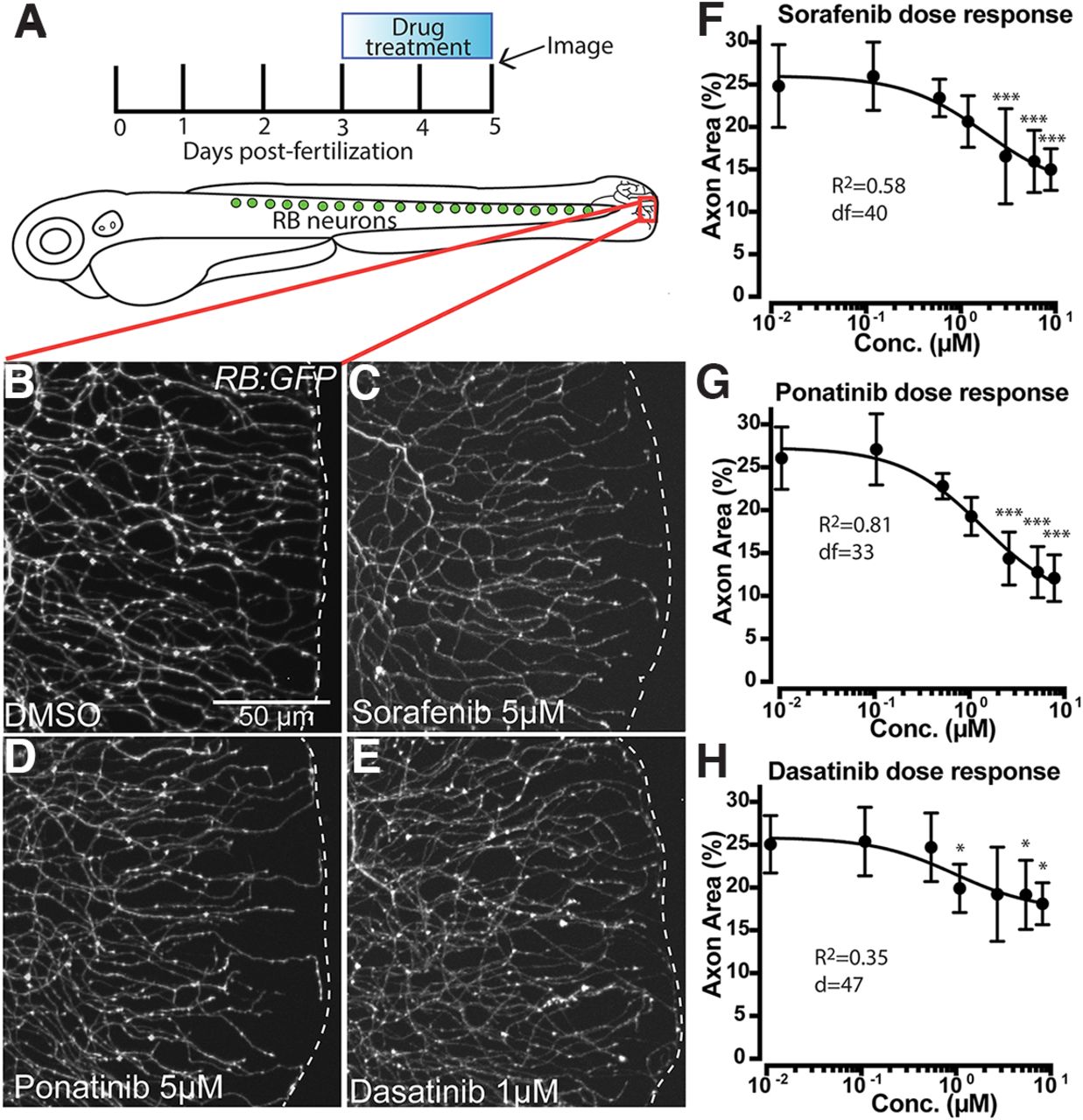
Fig. 1 Treatment with MKIs induces loss of cutaneous axon density in zebrafish caudal tail. A, Diagram of RB neurons, drug treatment and imaging paradigm, and imaging location (red box). B–E, Live images of somatosensory axons at the caudal tail tip labeled by RB:GFP after 48 h of treatment. Several MKIs produced significant loss of cutaneous axon density (white dashes indicate tail edge). F–H, Dose-dependent loss of distal axon density induced by (F) sorafenib, (G) ponatinib, and (H) dasatinib treatment, analyzed by one way ANOVA with post hoc Dunnett's test versus the lowest dose. Error bars represent SEM; *p < 0.05, ***p < 0.001.