
|
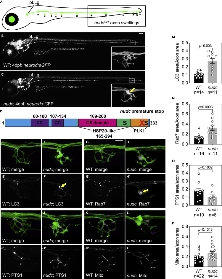
Fig. 1 NudC loss of function causes axon terminal swellings in the long axons of the zebrafish posterior lateral line (pLL) (A) Schematic of a larval zebrafish at 4 dpf. The pLL ganglion (pLLg) is indicated. Asterisks point to axon terminals in the pLL system. The posterior half of the trunk is where pLL axon terminal swellings are clearly observed in nudc mutants (boxed). (B and C) Wild type and nudc TgBAC(neurod:egfp)nl1 larva at 4 dpf. Insets show a magnified view of a distal axon terminal. Arrow points to axon terminal swelling in nudc mutants. (D) Diagram of NudC domains, including Plk1 phosphorylation sites at S274 and S326 (S) and the premature stop site in the nudcnl21 line (red X). Also shown are the coiled coil (CC), HSP20-like, and CS domains. (E–L) Localization of autophagosomes (E, F), late endosomes (G, H), peroxisomes (I, J), and mitochondria (K, L) in wild type and nudc axon terminals. Arrows in (F’) and (H’) indicate the accumulation of LC3+ and Rab7+ vesicles in nudc mutant axon terminals. (M−P) Average organelle load (organelle area/axon area) in axon terminals (Wilcoxon rank sum). Data are expressed as mean ± S.E.M. Sample sizes indicated. Scale bar = 100 μm in B and C; 10 μm in E–L.