
|
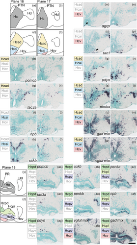
Fig. 26 Subregions in the caudal zone of the periventricular hypothalamus (Hc). (a), (b) The anterior part of Hc (gray) in the zebrafish brain atlas of Wullimann et al. (1996). (c), (d) The anterior part of Hc can be divided into three subregions: the anterodorsal (Hcad, yellow), anterointermediate (Hcai, blue), ventral (Hcv, pink) part of Hc. (e)–(h) Neuropeptides such as pomcb and tac3a are solely expressed in the Hcad. (i)–(l) Neuropeptides such as npb and cckb are expressed exclusively in the Hcai. (m)–(p) agrp and tac1 are only expressed in the Hcv. (q)–(t) Neuropeptides such as pdyn and penka are expressed across multiple subregions. (u)–(x) gad mix and vglut mix are expressed in all three subregions. (y) Posterior part of Hc (gray) in the zebrafish brain atlas of Wullimann et al. (1996). (z) The posterior part of Hc can be divided into four subregions: the posterodorsal (Hcpd, green), posterointermediate (Hcpi, light green), posteroventral (Hcpv, purple), and ventral (Hcv, pink) part of Hc. (aa), (ad), (ag) Neuropeptides such as pomcb, tac3a, and pdyn are solely expressed in the Hcpd. (ab), (ae) cckb and penkb are expressed in the Hcpd and Hcpv. (ah) vglut mix is expressed in Hcpd, Hcpv, and Hcv. (ac), (af) penka and npb are expressed in the Hcpd, Hcpi, and Hcpv. (ai) gad mix is expressed in all regions. Arrowheads indicate the location of weak signals. Scale bar = 100 μm.