
|
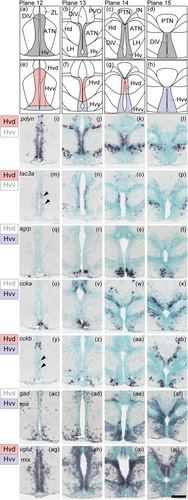
Fig. 24 Subregions in the ventral zone of the periventricular hypothalamus (Hv). (a)–(d) The Hv (gray) in the zebrafish brain atlas of Wullimann et al. (1996). (e)–(h) The Hv can be divided into two subregions: the ventral part of Hv (Hvv, purple) and the dorsal part of Hv (Hvd, pink). (i)–(x) Neuropeptides such as pdyn and tac3a are expressed only in the Hvd, whereas agrp and ccka are expressed only in the Hvv. (y)–(ab) cckb is expressed in both the Hvd and Hvv. (ac)–(aj) gad mix is predominantly expressed in the Hvv and the vglut mix is more densely expressed in the Hvd. Arrowheads indicate the locations of weak signals. Scale bar = 100 μm.