
Fig. 23
- ID
- ZDB-IMAGE-240620-140
- Publication
- Hiraki-Kajiyama et al., 2024 - An atlas and database of neuropeptide gene expression in the adult zebrafish forebrain
- All Figures
- Figures for Hiraki-Kajiyama et al., 2024
|
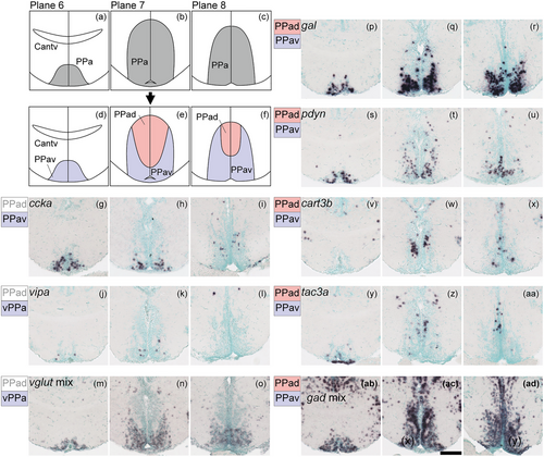
Fig. 23 Subregions in the anterior part of the parvocellular preoptic nucleus (PPa). (a)–(c) The PPa (gray) in the zebrafish brain atlas of Wullimann et al. (1996). (d)–(f) The PPa can be divided into two subregions: the ventral part of PPa (PPav, pink) and dorsal part of PPa (PPad, purple). (g)–(o) Neuropeptides such as ccka and vipa and vglut mix are exclusively expressed in the PPav. (p)–(u) gal and pdyn are expressed in both the PPad and PPav, but predominantly in the PPav. (v)–(aa) cart3b and tac3a are also expressed in the PPav and PPad but predominantly in the PPad. (ab)–(ad) gad mix is expressed in both the PPad and PPav. Scale bar = 100 μm.