
Image
|
Figure Caption
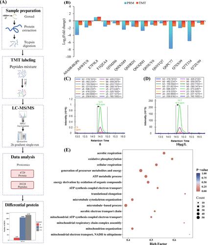
Fig. 3 Proteomic analysis in male zebrafish testes after exposure to 10 μg/L of BTBPE. (A) Schematic diagram of TMT-labeling proteome analysis. (B) The Log2 (fold change) of PRM results for TMT-based proteomic. (C, D) extracted-ion chromatograms for the representative peptide from Q7SX99 (fumarate hydratase protein) under 0 and 10 μg/L BTBPE treatments. (E) Significantly enriched GO pathways in biological process of DEPs of whole proteome.
Acknowledgments
This image is the copyrighted work of the attributed author or publisher, and
ZFIN has permission only to display this image to its users.
Additional permissions should be obtained from the applicable author or publisher of the image.
Full text @ Env. Sci. Tech.