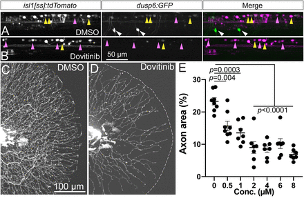
Fig. 7 Dovitinib induces RB axon loss and reduction in Fgf signaling similar to SU5402 treatment. A,B, Lateral view of isl1[ss]:tdTomato; dups6:eGFP dorsal neurons in live 4 dpf larvae. Compared to DMSO-treated controls (A), short-term (7 h) dovitinib treatment (B) induced a substantial loss of Fgf signaling-dependent GFP expression. C,D, Lateral view of RB:GFP larval caudal tails at 5 pf following 48 h DMSO (C) or dovitinib (D) treatment. Dovitinib treatment led to major axon loss similar to genetic and pharmacological loss of Fgfr signaling. E, Quantification of dose-dependent effect of dovitinib on tail axon density. DMSO = 23.3 ± 1.1%, 0.5 μM = 15.7 ± 1.5%, 1 μM = 13.5 ± 1.3%, 2 μM = 9.0 ± 1.8%, 4 μM = 8.5 ± 1.0%, 6 μM = 10.2 ± 1.5%, 8 μM = 6.9 ± 0.6%, analyzed by one-way ANOVA with post hoc Tukey's test (F = 19.61, p < 0.0001). Yellow arrowheads = EGFP+ RB neurons, magenta arrowheads = EGFP− RB neurons, white arrowheads = EGFP+ motor neurons.
Image
Figure Caption
Acknowledgments
This image is the copyrighted work of the attributed author or publisher, and
ZFIN has permission only to display this image to its users.
Additional permissions should be obtained from the applicable author or publisher of the image.
Full text @ J. Neurosci.

