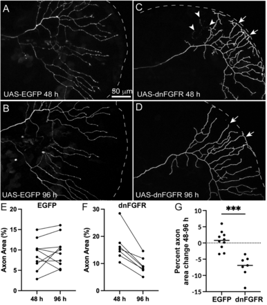
Fig. 6 Cell autonomous loss of Fgf signaling in RB neurons induces loss of axon density. A–D, Live-images of RB axons in the caudal tail mosaically expressing UAS-EGFP or UAS-dnfgfr1-EGFP in RB:RFP larvae from 48 hpf (A,C) to 96 hpf (B,D). E,F, Quantification of axon area % from 48 to 96 hpf from individual larvae injected with UAS-EGFP (C) or UAS-dnfgfr1-EGFP. Arrowheads mark sites of axon degeneration, whereas arrows mark axon terminals that lose elaboration over time. EGFP = 48 h = 8.83 ± 1.2%, 96 h = 9.6 ± 1.6%, p = 0.42, paired t test. dnfgfr = 16.5 ± 2.2%, 9.3 ± 1.2%, p = 0.001, paired t test. G, Comparison of change in axon area % of EGFP versus dnFgfr1-expressing RB axons from 48 to 96 hpf. EGFP = 0.7 ± 0.9%, dnFGFR = −7.7 ± 0.9, p = 0.0001, unpaired t test.
Image
Figure Caption
Acknowledgments
This image is the copyrighted work of the attributed author or publisher, and
ZFIN has permission only to display this image to its users.
Additional permissions should be obtained from the applicable author or publisher of the image.
Full text @ J. Neurosci.

