IMAGE

Figure 3

ID
ZDB-IMAGE-240429-132
Source
Figures for Veroni et al., 2024
Image
Figure Caption

Figure 3

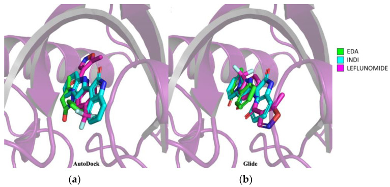
Prediction of EDA–AHR binding mode by molecular docking. (a) Superimposition of docking results on AHR of EDA in green, INDI in cyan and leflunomide in magenta using Autodock 4 software. (b) Superimposition of docking results on AHR of EDA in green, INDI in cyan and leflunomide in magenta using Glide software. In the 3D structures of the docked compounds, the oxygen atom is represented in red, nitrogen in blue while the fluorine atoms of leflunomide are represented in light blue. The image was edited using BioRender.com (https://www.biorender.com/ accessed on 3 April 2024).

Acknowledgments
This image is the copyrighted work of the attributed author or publisher, and ZFIN has permission only to display this image to its users. Additional permissions should be obtained from the applicable author or publisher of the image. Full text @ Biomolecules