
|
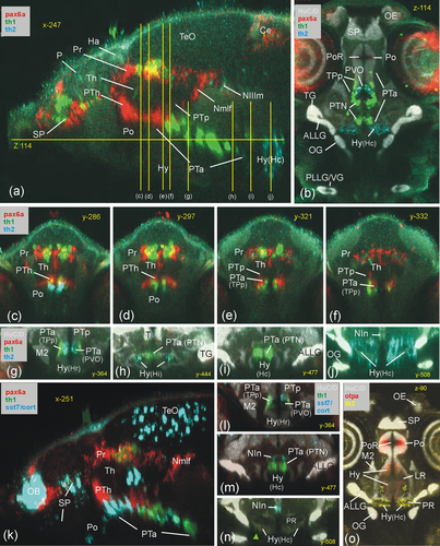
Fig. 9 Basal prosomere P3 zebrafish gene expression (anterior part of posterior tuberculum, PTa). Sagittal (a), horizontal (b) and transverse sections (c-j) characterize dopamine system (mesodiencephalic dopamine complex) and additional expression of sst7 (k: sagittal; l and n: consecutive transverse sections) and of otpa (o: sagittal). ALLG anterior lateral line ganglion; Ce, cerebellum; Ha, habenula; Hy, basal hypothalamus; Hy(Hc), caudal periventricular hypothalamic zone; Hy(Hi), intermediate periventricular hypothalamic zone; Hy(Hr), rostral periventricular hypothalamic zone; LR, lateral ventricular recess of periventricular hypothalamus; M2, early migrated posterior tubercular area (preglomerular complex); NIn, interpeduncular nucleus; Nmlf, region of the nucleus of the medial longitudinal fascicle; NIIIm, oculomotor nucleus; OB, olfactory bulb; OE, olfactory epithelium; OG, octaval ganglion; P, pallium; PLLG, posterior lateral line ganglion; Po, preoptic area; poc, postoptic commissure; PoR, preoptic ventricular recess; Pr, pretectum; PR, posterior ventricular recess of periventricular hypothalamus; PTa, anterior part of posterior tuberculum; PTh, prethalamus; PTN, posterior tuberal nucleus; PTp, posterior part of posterior tuberculum; PVO, paraventricular organ; SP, subpallium; T, midbrain tegmentum; TeO, tectum opticum; TG, trigeminal ganglion; TPp, periventricular posterior tubercular nucleus; VG, vagal ganglion.