
|
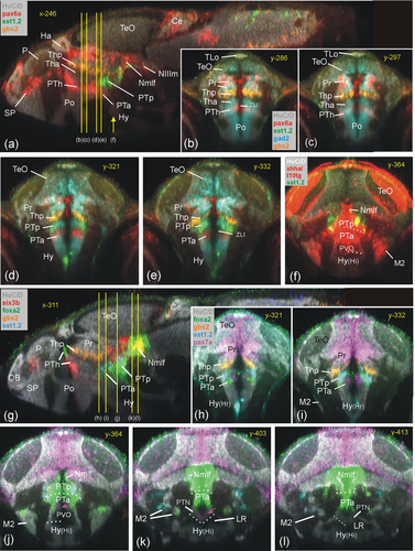
Fig. 6 Basal prosomere P2 zebrafish gene expression (posterior part of posterior tuberculum, PTp). Sagittal (a) and five consecutive transverse (b–f) sections emphasize diagnostic sst1.2 expression in PTp (additionally using gad2 and surrounding pax6a/gbx2 expression; B–E). (f) Basal plate marker shha in all three prosomere divisions including PTp (note overlap of shha with sst1.2). Sagittal (g) and five consecutive transverse sections (h–l) show PTp within basal diencephalon using basal plate marker genes foxa2 and pax7a together with sst1.2. Note six3b expression in Nmlf overlapping with foxa2 (g) and expression of foxa2 (j) and sst1.2 (k) as well as shha (f) in M2 (see text). Ce, cerebellum; Ha, habenula; Hy, basal hypothalamus; Hy(Hr), rostral periventricular hypothalamic zone; Hy(Hi), intermediate periventricular hypothalamic zone; LR, lateral ventricular recess of periventricular hypothalamus; M2, early migrated posterior tubercular area (preglomerular complex); Nmlf, region of the nucleus of the medial longitudinal fascicle; NIIIm, oculomotor nucleus; OB, olfactory bulb; P, pallium; Po, preoptic area; Pr, pretectum; PTh, prethalamus; PTa, anterior part of posterior tuberculum; PTp, posterior part of posterior tuberculum; PVO, paraventricular organ; SP, subpallium; TeO, tectum opticum; TLo, torus longitudinalis; ZLI, zona limitans intrathalamica.