
|
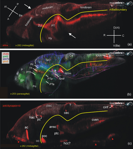
Fig. 2 Basic larval zebrafish brain organization. (a) Midsagittal section shows how longitudinal gene expression (example: sonic hedgehog a; shha) reflects on brain axes. (b) Parasagittal section shows exemplary gene expression for three diencephalic prosomeres (pax7a: pretectum, gbx2: thalamus, gad2 prethalamus). Note that single cells (see text) of basal pretectal prosomere projecting to brainstem and spinal cord were entered which constitute the nucleus of the medial longitudinal fascicle. (c) Midsagittal section shows brain commissures visualized using Synapsins (IHC). Note that optic nerve and chiasma are Synapsin negative (unlike optic tract; see text), but that there is Synapsin stain extraneous to the central nervous system (CNS) in roof of pharynx and gill chamber (arrowheads). For the commissural fiber staining marked with question marks in hypothalamus and cerebellum, see text. A, anterior; ac, anterior commissure; Al, alar; ansc, ansulate commissure; Ba, basal; C, caudal; Ce, cerebellum; cec, cerebellar commissure; cinf, commissura infima of Haller; ctec, tectal commissure; cven, ventral rhombencephalic commissure; D, dorsal (in hindbrain identical to alar); hac, habenular commissure; hcc, caudal hypothalamic commissure; Hy, hypothalamus; Nmlf, nucleus of the medial longitudinal fascicle; NIIIm, oculomotor nucleus; NXm, vagal motor nucleus; oc, optic chiasma; P, posterior (in A); P, pallium (in B); pc, posterior commissure; Po, preoptic region; poc, postoptic commissure; Pr, pretectum; PT, posterior tuberculum; ptc, posterior, tubercular commissure; PTa, anterior part of posterior tuberculum; PTh, prethalamus; PTp, posterior part of posterior tuberculum; R, rostral; SP, subpallium; T, midbrain tegmentum; TeO, tectum opticum; Th, thalamus; V, ventral (in hindbrain identical to basal); ZLI, zona limitans intrathalamica.