IMAGE

FIGURE 7

ID
ZDB-IMAGE-240323-33
Source
Figures for Wang et al., 2024
Image
Figure Caption

FIGURE 7

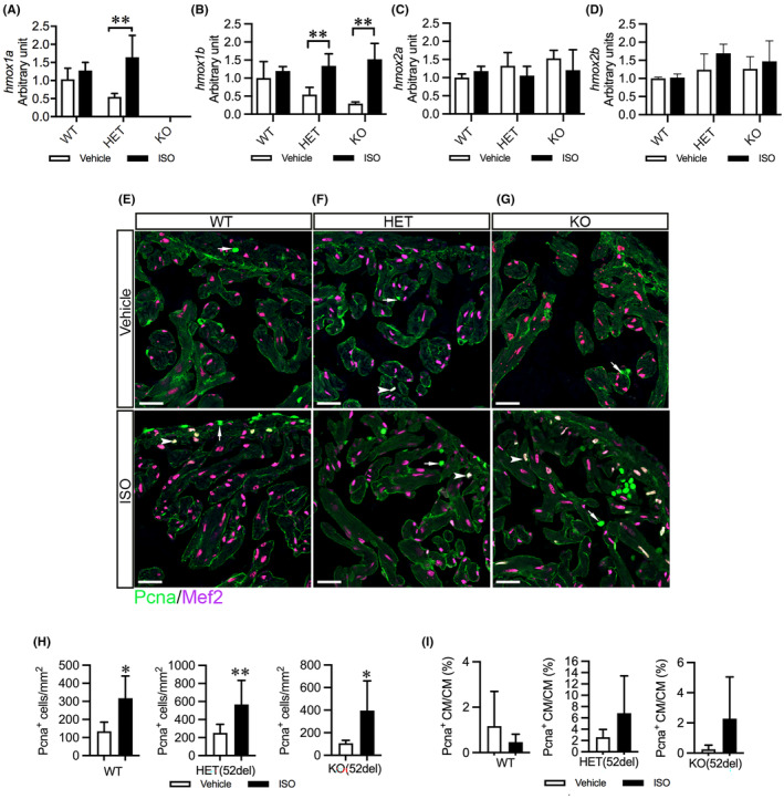
ISO upregulates cardiac hmox1 paralogs and increases cardiac cell proliferation in HET(52del) adults. (A–D) RT‐qPCR analyses of the expression of hmox1 homologues in ISO‐induced zebrafish hearts compared to vehicle‐treated controls. The results are presented as fold change compared to vehicle‐treated WT hearts. WT, vehicle n = 6, ISO n = 4; HET(52del), vehicle n = 6, ISO n = 4; KO(52del), vehicle n = 5, ISO n = 4. (E–G) Representative immunohistochemical staining of Pcna (in green) and Mef2 (in meganne) in ventricular sections from vehicle‐ or ISO‐treated zebrafish in each genotypic group. Arrow indicates Pcna+ non‐cardiomyocytes. Arrowhead indicates Pcna+ cardiomyocytes. (H, I) Quantification of Pcna+ cardiac cells per mm2 ventricular area (H) and Pcna+ cardiomyocytes relative to cardiomyocytes (I). Three hearts from each genotypic group were chosen. Two sections from each heart were stained. The number of stain‐positive signals for Pcna and Mef2 from ventricular areas was quantified with ImageJ. Data are presented as mean ± SD. Two‐sample t‐test. *p < 0.05, **p < 0.01.

Acknowledgments
This image is the copyrighted work of the attributed author or publisher, and ZFIN has permission only to display this image to its users. Additional permissions should be obtained from the applicable author or publisher of the image. Full text @ J. Cell. Mol. Med.