
|
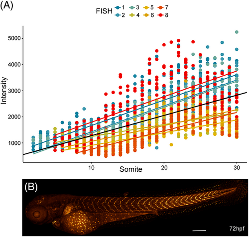
Fig. 7 tgfbr3 promoter has higher activity in slow muscle fibers of the posterior trunk somites. (A) The linear regression shows an increase in the normalized mCherry fluorescence intensity with respect to somite location. Each point in the plot represents the value obtained for single nuclei after normalizing with Hoechst staining as described in the text. Intensity fluorescence from eight nuclei were measure per somite, in eight different embryos at 72 hpf. Individual slopes are color-coded, and the overall regression is in black. (B) Representative image of the A-P gradient expression gradient in 72 hpf Tg(tgfbr3:nls-mCherry) embryo.