
|
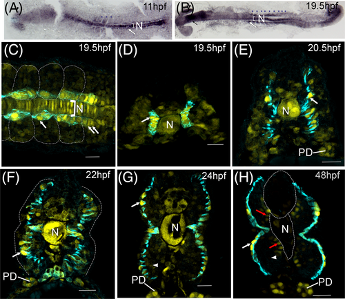
Fig. 6 tgfbr3 promoter is upregulated in slow muscle and marks adaxial cells during their radial migration. (A, B) Immunohistochemistry for mCherry in flat mounted transgenic zebrafish embryos. High mCherry expression is detected in the notochord (N), in somitic mesoderm (indicated by blue asterisks) with higher signal in adaxial cells (white arrows) from presomitic and somitic mesoderm at 3 somite (11 hpf) and 19 somite stages (19.5 hpf). Expression is also observed in the eye (black arrowhead) and head (black arrow). (C-H) immunofluorescent staining of mCherry (yellow) and F59 (cyan). Dorsal view (panel C) of an embryo at 19.5 hpf (somites 16-19). Adaxial cells were labeled with F59 (cyan), mCherry is observed with higher fluorescence in the nuclei of adaxial cells from the somitic mesoderm (white arrow) and adaxial cells from presomitic mesoderm (double white arrows). Cells in the notochord (N) express high levels of mCherry. Transverse sections (panels D-H) in the caudal trunk (somites 14-17) show mCherry in the nuclei of adaxial cells (white arrows), which are revealed by the F59 staining (cyan). This monoclonal antibody marks the adaxial cells during their radial migration from 19.5 to 24 hpf (panels D-G) all the way to the somite surface where they become slow fibers (panel H). mCherry expression in notochord (N) and somitic fast muscle decreases gradually (white arrowheads in panels G and H), except in the fast fibers in contact with the notochord and neural tube (red arrows), while in the nuclei of superficial slow muscle fibers the mCherry expression increases (white arrows). The expression in pronephric ducts (PD) increases as development proceeds. In dorsal views, embryos are oriented with head to the left. Scale bars, 20 μm.