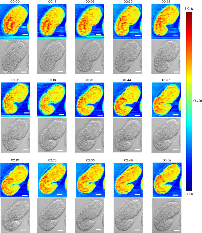
IMAGE

Fig. 5

ID
ZDB-IMAGE-231211-56
Source
Figures for Yang et al., 2023
Image
Figure Caption

Fig. 5 Time-lapse pulsed-SBS imaging of C. elegans embryo development.

Brillouin shift (top) and brightfield (bottom) images over a 3-h time span and at 13-min time interval (representative of n = 3 embryos). The pixel steps in x and y directions are 0.5 µm and the pixel time is 20 ms (total time per image 288 s). Scale bars in all the images are 10 µm. The embryo at the start of the time lapse is at the late ‘ball-stage’, slightly before the bean stage, or approximately at 300 min post-fertilization (mpf). Imaging time lapses presented here conclude at ~480 mpf, when embryos start their first spontaneous muscle contractions, known as ‘twitching’. Embryonic axes: top right is anterior, bottom left is posterior, right is ventral and left is dorsal.

Acknowledgments
This image is the copyrighted work of the attributed author or publisher, and ZFIN has permission only to display this image to its users. Additional permissions should be obtained from the applicable author or publisher of the image. Full text @ Nat. Methods