
|
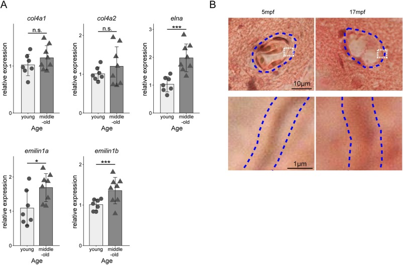
Fig. 7 Aging affects the expression of vessel wall ECM-related genes. (A)The results of qRT–PCR analysis of gene expression. Each bar represents the average of the relative expression. The circles indicate the individual expression levels in young (4–5 mpf) fish (n = 7) and the triangles indicate the same in middle-old-aged (14–15 hpf) fish (n = 8). n.s. represents no significant difference; *, p < 0.05, ***; p < 0.001 (Welch's t-test). (B) Elastic fiber staining in the telencephalon of young and middle-old-aged fish. The results of resorcin-fuchsin and Van Gieson's staining in 5mpf and 17 mpf fish telencephalon section. Black purple-colored elastic fiber staining was reduced age-dependently. Blue dotted lines show the blood vessel in upper panels. Lower panels show the enlarged view of the white dotted line area in the upper panels. Blue dotted lines in lower panels indicate the vessel wall. Scale bars show 10 μm and 1 μm, respectively. (For interpretation of the references to colour in this figure legend, the reader is referred to the web version of this article.)