IMAGE

Figure 8

ID
ZDB-IMAGE-231016-32
Source
Figures for Chen et al., 2023
Image
Figure Caption

Figure 8

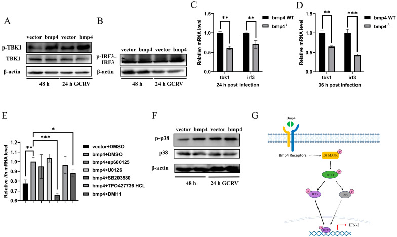
Bmp4 increases Tbk1-Irf3 antiviral signaling via p38 MAPK pathway. (A,B) Immunoblot analysis of phosphorylated (p-) Tbk1 (A) and Irf3 (B) was performed 24 h after transfection of 2 μg of bmp4 or empty vector in EPC cells, followed by infection with or without GCRV for 24 h. (C,D) Expression of tbk1 and irf3 mRNA from WT or bmp4−/− zebrafish larvae challenged with GCRV. (E) Expression of EPC ifn mRNA 24 h after transfection of bmp4 (2 μg) in EPC cells, followed by treatment with SB203580, SP600125, U0126, DMH1 and TP0427736 HCl for 24 h. (F) Representative Western blot analysis in p-p38 MAPK expression in bmp4-overexpressed EPC cells. (G) Schematic illustration of the regulation of Bmp4 in the antiviral immune response. The Bmp4 promotes phosphorylation of Tbk1 and Irf3 to induce the expression of ifn through p38 MAPK pathway. The expression of zebrafish actb1 or EPC actin was used as an internal control for the qRT-PCR. Data were from three independent experiments and were analyzed through Student’s t-test (two-tailed) for comparison of two groups or one-way ANOVA followed by Games-Howell post hoc tests for comparison of multiple groups. All data are presented as mean ± SD (* p < 0.05, ** p < 0.01, *** p < 0.001).

Acknowledgments
This image is the copyrighted work of the attributed author or publisher, and ZFIN has permission only to display this image to its users. Additional permissions should be obtained from the applicable author or publisher of the image. Full text @ Int. J. Mol. Sci.