IMAGE

Fig. 3

ID
ZDB-IMAGE-231002-85
Source
Figures for Tsai et al., 2023
Image
Figure Caption

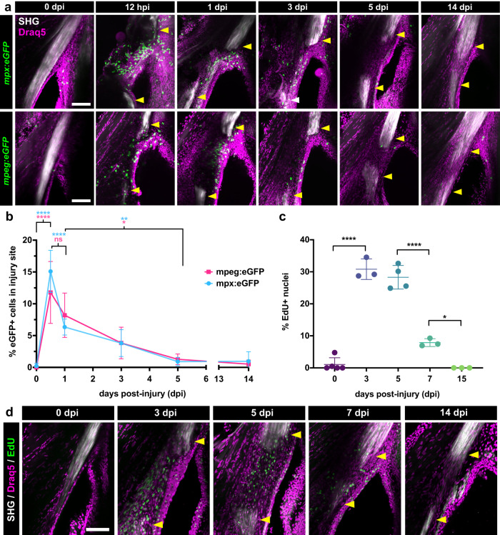
Fig. 3 Tendon injury triggers a rapid innate immune response followed by a wave of cellular proliferation.

a 2-photon imaging of regenerating tendons from mpx:eGFP (top row) or mpeg1:eGFP (bottom row) zebrafish at different time points post-injury to examine neutrophil and macrophage dynamics, respectively. SHG signal is overlaid along with a Draq5 nuclear counterstain. Yellow arrowheads denote severed tendon ends. Scale bar, 100 µm. b Quantification of the percentage of mpx:eGFP+ and mpeg1:eGFP+ cells out of total Draq5+ cells in the injury site during the first 2 weeks post-injury. One-way ANOVA analysis was employed for statistical analysis with Tukey’s multiple comparison tests between different time points. Sample sizes were as follows: mpx:eGFP – 0, 1, 14 dpi: N = 4; 0.5, 3, 5 dpi: N = 5; mpeg1:eGFP – 0.5 dpi: N = 6; 0, 1, 3 dpi: N = 5; 5 dpi: N = 4; 14 dpi: N = 3. ****p < 0.0001, **p < 0.01, *p < 0.05. c Quantification of the percentage of EdU+ cells out of total Draq5+ cells in the injured area at 0, 3, 5, 7, and 15 dpi. One-way ANOVA analysis was employed for statistical analysis with Tukey’s multiple comparison tests between time points. Sample sizes were as follows: 0 dpi: N = 5; 3, 7, 15 dpi: N = 3; 5 dpi: N = 4. ****p < 0.0001, *p < 0.05. d Representative 2-photon time course imaging of EdU+ cells (in green) at different time points post-injury. SHG signal is overlaid with Draq5 nuclear staining. Yellow arrowheads denote severed tendon ends. Scale bar, 100 µm. All error bars in graphs denote the standard deviation.

Acknowledgments
This image is the copyrighted work of the attributed author or publisher, and ZFIN has permission only to display this image to its users. Additional permissions should be obtained from the applicable author or publisher of the image. Full text @ NPJ Regen Med