IMAGE

Figure 1.

ID
ZDB-IMAGE-231002-383
Source
Figures for Brondolin et al., 2023
Image
Figure Caption

Figure 1.

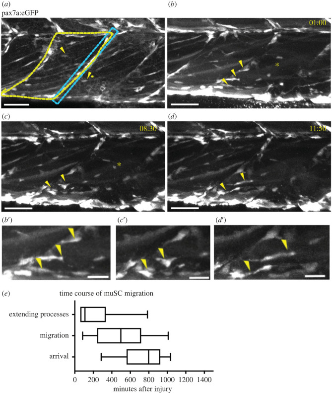
MuSCs show a transition in shape as they move from the myoseptum towards the site of injury (asterisk). GFP + muSCs (arrowheads) in uninjured 7 dpf pax7a:egfp larvae are located at the myoseptum (blue box) or in the myotome (yellow outline, a). In the presence of an injury GFP + muSCs at the vertical borders of the myoseptum extend processes into the myotome (b′, arrowheads) and undergo extensive movement along the myoseptum (b). They then stabilize a process extending into the myotome (c′, arrowheads) and detach from the myoseptum (c). Subsequently the GFP + muSCs migrate towards the site of injury, extending processes in multiple directions (d′, arrowheads) as they migrate (d). Quantification of cell responses to injury reveal three phases: extension of cell process, migration and arrival at the injury site (e, n = 28 cells from 6 animals). Scale bar 50 µm (a–d), 20 µm (b′–d′).

Acknowledgments
This image is the copyrighted work of the attributed author or publisher, and ZFIN has permission only to display this image to its users. Additional permissions should be obtained from the applicable author or publisher of the image. Full text @ Open Biol.