
|
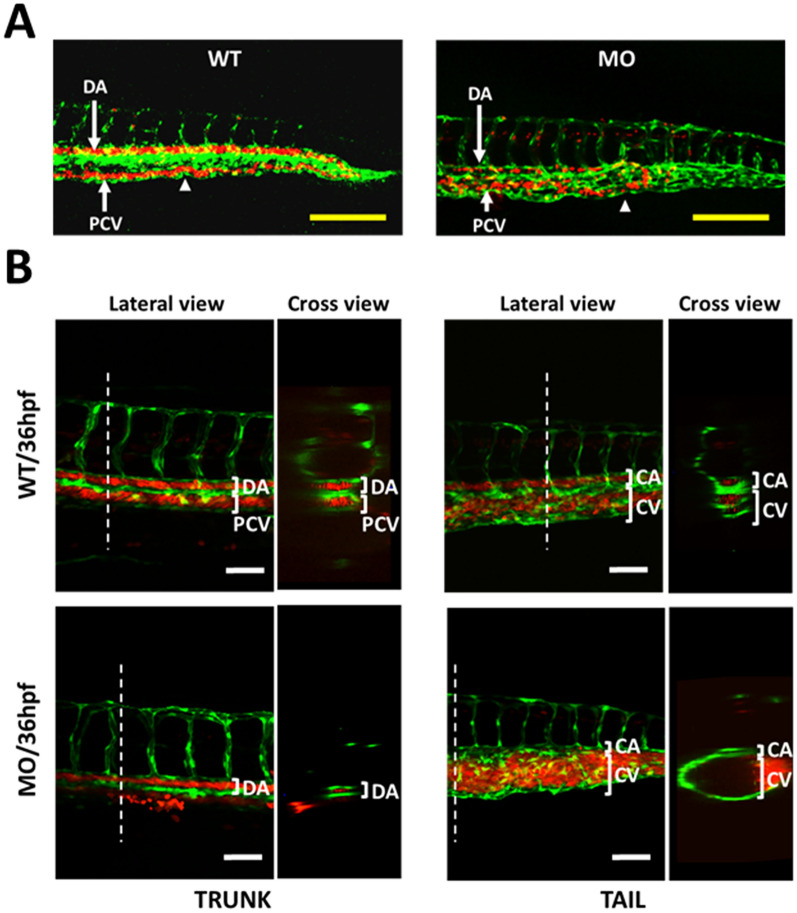
Fig. 1
Effects of YULINK knockdown on the vasculature in zebrafish embryos. A The double transgenic zebrafish Tg (fli1:EGFP; gata1:DsRed) displayed fli1 promoter-derived green fluorescent for endothelial cells and gata1 promoter-derived red fluorescent for erythrocytes. Arrows indicate the anus of the embryo. Yellow scale bars indicate 200 μm. B The confocal images of the trunk and caudal vasculature at 36 hpf are shown with their lateral and cross-section views. The white dotted lines indicate the position where the cross-sections were taken. DA dorsal aorta, PCV posterior cardinal vein, CA caudal artery, CV caudal vein. White scale bars indicate 50 μm