
|
Fig. 3
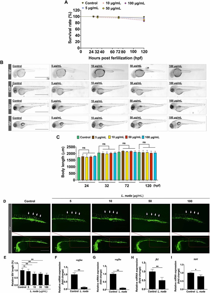
Fig. 3. Representative and quantitative results of L. nuda extract treatment on zebrafish embryo development and inhibition of ISV growth. (A) The relative survival rate of zebrafish embryos showed no cytotoxicity of L. nuda (5–100 μg/mL) treatment (24–120 hpf). (B, C) The body length of zebrafish embryos was measured after L. nuda (5–100 μg/mL) treatment. (D) Lateral assessment of Tg (fli1: EGFP) zebrafish embryos at 24 h postfertilization (hpf) revealed vessel destruction following L. nuda treatment. (E) Relative ISV length was significantly reduced after L. nuda treatment. The white arrow indicates the ISV location. (F-I) The depletion of mRNA levels of vegfaa, vegfba, kdrl, and flt1 following L. nuda treatment (10 μg/mL) as analyzed by qPCR. * p < 0.05 and * * p < 0.001; Scale bar = 100 µm.