
Image
|
Figure Caption
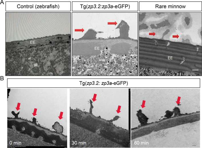
Fig. 3
Gain of partial adhesiveness in transgenic zebrafish eggs
A: TEM images showing structures in outer layer of 0 hpf fertilized eggs from WT zebrafish, Tg(zp3.2: zp3a-eGFP) line, and rare minnow. Red arrows indicate electron-dense projections. Bar: 2 μm. B: TEM images showing budlike structures on outer layer of transgenic eggs at indicated time points. Red arrows indicate electron-dense projections. Bar: 1 μm.
Acknowledgments
This image is the copyrighted work of the attributed author or publisher, and
ZFIN has permission only to display this image to its users.
Additional permissions should be obtained from the applicable author or publisher of the image.
Full text @ Zool Res