IMAGE

FIG. 2.

ID
ZDB-IMAGE-230625-11
Source
Figures for Zhang et al., 2023
Image
Figure Caption

FIG. 2.

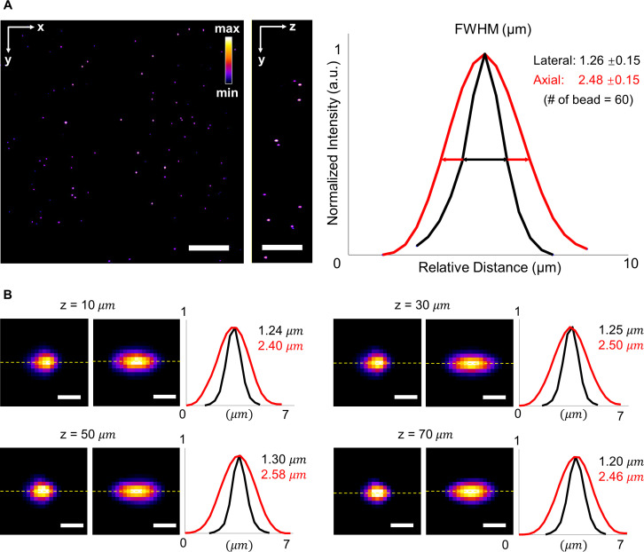
Full width at half maximum (FWHM) of beads captured by LSFM at various depths. (a) Raw data of fluorescent beads in a volume of ∼300 × 300 × 75 μm3 in the left panel. The average lateral and axial resolutions of the LSFM are 1.26 ± 0.15 μm and 2.48 ± 0.15 μm, respectively. Normalized intensity is shown in Fire pseudo color. (b) Cross section images of beads at different depths ranging from 0 to 75 μm. Representative lateral and axial resolutions at different depths were measured along the dash lines. Scale bars: (a) 50 and (b) 2 μm.

Acknowledgments
This image is the copyrighted work of the attributed author or publisher, and ZFIN has permission only to display this image to its users. Additional permissions should be obtained from the applicable author or publisher of the image. Full text @ APL Bioeng