
|
Fig. 5 Molecular defects in craniofacial development and chondrocyte morphology in Shoc2 depleted embryos.
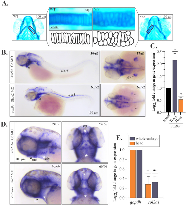
(A) Flat mounts of WT and shoc2(Δ22) mutant 6 dpf larvae stained with Alcian blue. The lower panels show individual chondrocytes outlined in black, highlighting the differences in cell morphology of the ceratohyal cartilage. (B) Lateral and dorsal views of control and shoc2 morphant larvae show the sox9a expression of 2 dpf larvae. Asterisks show areas of altered sox9a expression in the control and shoc2 morphants. (C) Total RNA was extracted from the dissected trunk of 3 dpf control and shoc2 morphant larvae. The levels of sox9a mRNA expression were quantified by qPCR. gapdh is a control mRNA. The data are presented as the Log2fold change of the mRNA levels in morphant larvae normalized to control. The results represent an average of three biological replicas. Error bars indicate means with SEM. ∗p<0.05, ∗∗p<0.01, ∗∗∗p<0.001 (Student’s t-test). (D) Lateral and dorsal views of control and shoc2 morphant embryos show the col2a1 expression in 3 dpf larvae. Asterisks mark areas of reduced col2a1 expression in shoc2 morphants. (E) Total RNA was extracted from dissected 3 dpf control and Shoc2 morphant larvae and levels of col2a1 mRNA expression were quantified by qPCR. The data are presented as the Log2fold change of the mRNA levels in morphant larvae normalized to control. gapdh is a control mRNA. The results represent an average of three biological replicas. Error bars indicate means with SEM. ∗p<0.05, ∗∗p<0.01, ∗∗∗p<0.001 (Student’s t-test). pf: pectoral fin. mc: Meckel’s cartilage. cbs: ceratobranchials. ep: ethmoid plate.
Reprinted from Developmental Biology, 492, Norcross, R.G., Abdelmoti, L., Rouchka, E.C., Andreeva, K., Tussey, O., Landestoy, D., Galperin, E., Shoc2 controls ERK1/2-driven neural crest development by balancing components of the extracellular matrix, 156-171, Copyright (2022) with permission from Elsevier. Full text @ Dev. Biol.