
|
Fig. 3
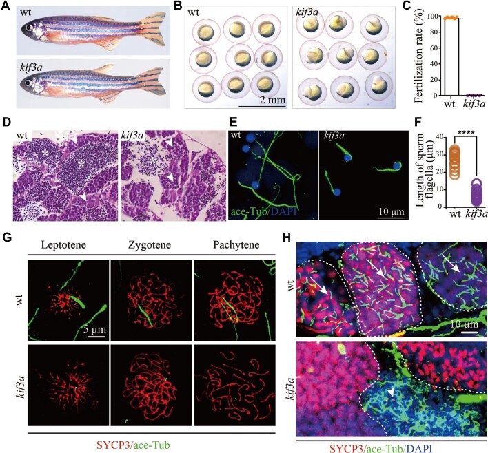
Phenotypes of germ cell-specific kif3a conditional knockout mutants. (A) External phenotypes of wild type and Tg(kop:cas9-UTRnanos3;U6:3×sgRNA-kif3a) double transgenic male fish. (B) The embryos produced from wild type female crossed with control or double transgenic male at 6 hpf. (C) Statistical results showing the percentages of fertilization rates of wild type and double transgenic males as indicated. (D) Histological analysis of testis of wild type and Tg(kop:cas9-UTRnanos;U6:3×sgRNA-kif3a) double transgenic fish. Arrowheads point to spermatocytes and asterisks indicate spermatozoa. (E) Confocal images showing sperm flagella in wild type and double transgenic fish labelled with anti-acetylated tubulin antibody (green). (F) Statistical analysis of the length of sperm flagella in wild type and double transgenic fish. (G) Confocal images showing cilia in primary spermatocytes of wild type and double transgenic fish. Spermatocyte cilia (green) were absent in the double transgenic fish. The stages of spermatocytes were distinguished by the staining of SYCP3 (red). (H) Staining of SYCP3 (red) and acetylated-tubulin (green) in the testis of wild type and double transgenic fish. Arrows indicate spermatocyte cilia, while arrowheads indicate the staining of abnormal sperm flagella in the mutant testis. ****P < 0.0001. wt, wild type.