
|
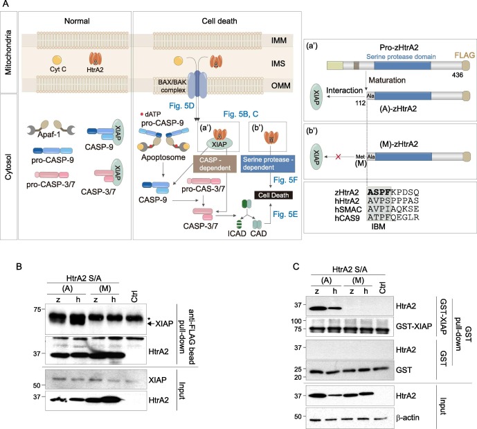
Fig. 5 Fig. 5. zHtrA2 induces CASP-independent/serine protease-dependent or CASP-dependent cell death. (A) Schematic representation of HtrA2-mediated cell death. Under normal conditions, HtrA2 and Cyt C are localized in the mitochondrial IMS. Pro-CASP-9 and pro-CASP-3/7 are inactive zymogen or proenzyme initiator and executioner CASPs, respectively. XIAP binds to and inhibits active CASP-9 and -3/-7. In response to cell death stimuli, BAX/BAK dimers generate pores within the OMM, causing MOMP and the non-selective release of Cyt C and HtrA2 into the cytosol (Fig. 5D). The released Cyt C binds to apoptotic protease activating factor 1 (Apaf-1) to form oligomeric apoptosomes, followed by activation of the CASP cascade to induce CASP-dependent cell death. (a′) and (b′) are described in more detail in the right panel. (B) Interaction of hXIAP and zHtrA2 through the exposed N-terminal XIAP that antagonizes IBM. After performing the anti-FLAG bead pull-down like Fig. 4B, the samples were probed with Abs against the indicated proteins. HtrA2 was detected by D-Tag Ab. The (M)-HtrA2 denotes the N-terminal Met (M) in HtrA2 preceding the IBM shown in Fig. 5A (b′). The asterisk indicates the IgH chain. (C) The interaction between zHtrA2 and XIAP. The interaction between proteins was verified with GST pull-down by incubating the GST proteins bound to Glutathione Sepharose 4B beads and the cell lysates containing the indicated HtrA2 proteins for 16 h at 4 °C. (D) Verification of mitochondrial release of HtrA2 into the cytosol via BAX-mediated MOMP. HEK293T cells were co-transfected with plasmids encoding C-terminal FLAG tagged pro-HtrA2 S/A and BAX, HtrA2 was immunostained with D-Tag Ab. The cells were treated with 20 μM pan-CASP inhibitors 6 h after transfection to maintain cell shapes. In parallel, whole-cell lysates (W) were separated into the mitochondrial (M) and cytosol (C) fractions, and probed with Abs for the indicated proteins. Voltage-dependent action channel (VDAC) and β-actin were used as mitochondrial (M) and cytosolic (C) markers, respectively. (E) The role of released zHtrA2 in promoting CASP-dependent cell death by inhibiting the XIAP-mediated inhibition of CASPs. For XIAP inhibition by HtrA2, GSH-bound GST-XIAP was incubated for 16 h at 4 °C with (A)-HtrA2 S/A pulled down with FLAG beads and incubated with the HEK293T S100 extract with Cyt C and dATP for 90 min at 30 °C. CASP-dependent cell death was determined by detection of active CASP-9/-3 and cleaved ICAD by IB. (F) Determination of serine protease-dependent cell death. HEK293T cells were co-transfected with plasmids encoding the indicated proteins; the plasmid expressing GFP was used as the transfection control. Ctrl indicates cells transfected only with the plasmid encoding GFP. At 16 h post-transfection, the GFP-positive (GFP + ) cells were visualized by fluorescence microscopy. The values on the y-axis were expressed as percentages (%) by dividing the number of round (dead, white arrow) GFP + cells by the total number of GFP + cells. The data are presented as means ± SEM, and the significance of the differences was calculated by the paired t-test using GraphPad Prism 8.
Reprinted from Gene, 819, Nam, M.K., Moon, J.M., Kim, G.Y., Min Kim, S., Rhim, H., The Novel Human HtrA2 Ortholog in Zebrafish: New Molecular Insight and Challenges into the Imbalance of Homeostasis, 146263, Copyright (2022) with permission from Elsevier. Full text @ Gene