
|
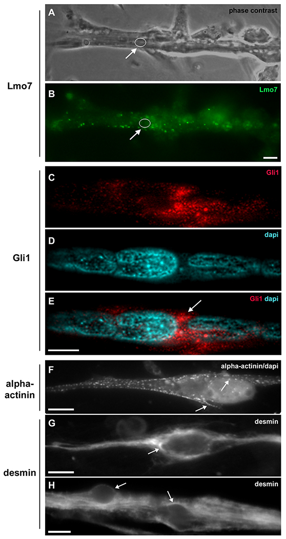
Fig. 3
Lmo7, Gli1, alpha-actinin and desmin are concentrated in the nuclear cloud in skeletal muscle fibers. Primary cultures of chick myogenic cells were labeled with antibodies against Lmo7 (B), Gli1 (C–E), alpha-actinin (F) and desmin (G–H), and with the nuclear dye DAPI (D–F). A 72-h chick multinucleated myotube was visualized under phase contrast microscopy (A) and under fluorescence microscopy to show the localization of Lmo7 (green, in B). Arrows in A and B point to Lmo7 distribution near the nuclear surface of a myotube. White open circles (in A and B) mark the region of one nucleus surrounded by Lmo7-positive aggregates. Scale bar in A = 10 μm. Gli-1 (red, in C and E) localizes at the perinuclear region of a 72-h multinucleated myotube (arrow in E). A merged image (with Gli1 and DAPI) is shown in E. Scale bar in E = 5 μm. The intermediate filament desmin and the sarcomeric protein alpha-actinin accumulate at the perinuclear region of mononucleated myoblasts (F and G) and multinucleated myotubes (H). A merged image of sarcomeric alpha-actinin and DAPI is shown in (F). Note the punctate distribution of alpha-actinin (arrows in F) and the continuous distribution of desmin filaments (arrows in G and H) in the juxtanuclear region of cells. Scale bars in F and H = 10 μm, and in G = 5 μm. n = 4 independent experiments.
Reprinted from Biochimica et biophysica acta. Molecular cell research, 1869, do Amaral, M.J., de Andrade Rosa, I., Andrade, S.A., Fang, X., Andrade, L.R., Costa, M.L., Mermelstein, C., The perinuclear region concentrates disordered proteins with predicted phase separation distributed in a 3D network of cytoskeletal filaments and organelles, 119161, Copyright (2022) with permission from Elsevier. Full text @ BBA Molecular Cell Research