
|
Fig. 1
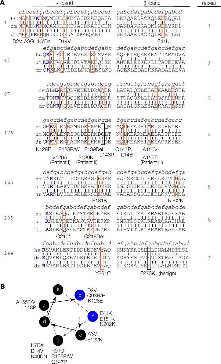
TPM2 residues associated with pathogenic variants are conserved. (A) TPM2 conservation and sequence structure. TPM2 protein sequence divided into 7 quasi-repeats, split into α- and β-bands as described before (26). Actin-binding residues are colored blue. Vertical black lines show identical residues between the human (hs), Drosophila (dm), or zebrafish (dr) proteins; gray lines show similar residues. A total of 25 pathogenic TPM2 coding region variants have been reported (19). We identified 2 potentially novel variants (V129A and E139K) and 1 recurring variant (A155T) in patients with musculoskeletal birth defects. Variants affecting conserved residues are boxed in red; nonconserved pathogenic variants are boxed in black. (B) Conserved pathogenic variants disproportionately cluster to a single topographical position. Diagram showing the 7–amino acid heptad (a–g) of the TPM2 coiled-coil, as described (). A subset of b and f residues binds actin (blue circles). There are 7 conserved pathogenic variants mapped to residues in position g.