
|
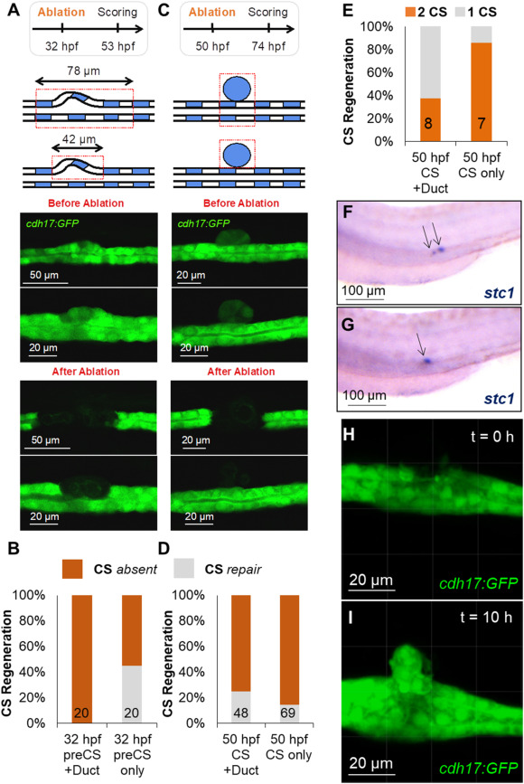
Fig. 3 Laser-ablated corpuscles of Stannius (CS) can partially regenerate.
(A) Laser ablation was used to destroy either the developing CS (preCS) together with the surrounding tubule (Duct) (78 μm), or only the developing CS (preCS) (42 μm) at 32 hpf. CS development was scored at 53 hpf. Top panel shows schematics of both setups. Middle panel shows the CS before ablation and the bottom panel shows the laser-ablated pronephric tubules. (B) Quantification of the experiments shown in (A). The numbers in the bars show the number of scored embryos. After the circumscribed 42 μm-laser ablation, GFP-positive material was detectable in 45% of the embryosthat likely represents regenerating CS. (C) Ablation of either CS plus the underlying tubule (CS + Duct), or selectively the CS (CS only) at 50 hpf. Scoring was done at 74 hpf. Top panel shows schematics of both setups. Middle panel shows the CS before ablation and the bottom panel shows the laser-ablated pronephric tubules. (D) Quantification of the experiments shown in (C). The numbers in the bars show the number of scored embryos. Some GFP-positive material was detectable in ca. 20%, likely representing newly formed CS. (E) Embryos with putative newly formed CS after ablation at 50 hpf were stained for stc1. In ca. 30% of the cases after CS + duct ablation and ca. 90% of the cases where only the CS was ablated two CS were detectable. (F) An example of embryo with two stc1 positive CS (arrows) after ablation as described in (E). (G) An example of embryo with one stc1 positive CS (arrows) after ablation as described in (E). (H) A still image from a time lapse of CS regeneration showing the pronephric tubule after CS ablation at 50 hpf. (I) A still image from a time lapse of CS regeneration showing the regenerated CS at 10 h post ablation as described in (H).
Reprinted from Developmental Biology, 481, Klingbeil, K., Nguyen, T., Fahrner, A., Guthmann, C., Wang, H., Schoels, M., Lilienkamp, M., Franz, H., Eckert, P., Walz, G., Yakulov, T.A., Corpuscles of Stannius development requires FGF signaling, 160-171, Copyright (2021) with permission from Elsevier. Full text @ Dev. Biol.