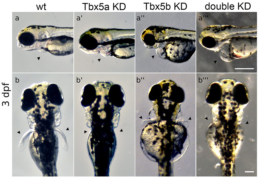
Fig. 1 Tbx5 knock-down embryos display heart and pectoral fin defects. (a-a’’’) Arrowheads point to position of heart. Views are lateral with anterior to the left. Scale bar=250μm. (a) Normal wt heart development. (a’) Tbx5a-knock-down embryos display edema and defects in heart looping. (a’’) Tbx5b-knock-down embryos display edema and defects in heart looping. (a’’’) Double knock-down embryos have linear hearts and edema, (b-b’’’) Arrowheads point to normal position of pectoral fins. Views are from dorsal aspect with anterior to the top. Scale bar=100μm. (b) Wt embryo showing both fins at 3 days post-fertilization (dpf). (b’) Tbx5a knock-down embryos lack pectoral fins, (b’’) Tbx5b knock-down embryos display small, misshapen fins. (b’’’) Tbx5a/Tbx5b double knock-down embryos do not form pectoral fins. Panels a, a’’, b and b’’ reproduced from Boyle Anderson and Ho (2018).
Reprinted from Developmental Biology, 481, Anderson, E.B., Mao, Q., Ho, R.K., Tbx5a and Tbx5b paralogues act in combination to control separate vectors of migration in the fin field of zebrafish, 201-214, Copyright (2021) with permission from Elsevier. Full text @ Dev. Biol.

