
Image
|
Figure Caption
Figure 5
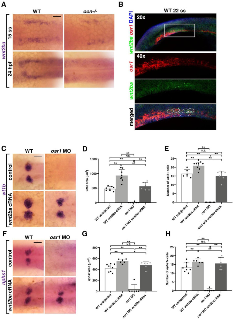
Figure 5. wnt2ba is sufficient for podocyte development downstream of osr1. (A) When wnt2ba was assessed at 15 ss, in ocn−/− and WT siblings, it was evident that staining was reduced in mutants. By 24 hpf, wnt2ba staining was almost completely absent, tracking with the loss of other podocyte markers in ocn−/−. Scale bar is 30 μm. (B) FISH experiments showed that osr1 and wnt2ba colocalized in cells at 22 ss. (C–H) Embryos were injected with osr1 MO and/or wnt2ba cRNA at the one-cell stage, and podocytes and the developing slit diaphragm were visualized at 24 hpf using wt1b and nphs1, respectively. Embryos injected with osr1 MO alone showed few podocyte or slit diaphragm cells, while embryos injected with wnt2ba cRNA alone had an increased podocyte area. Coinjected embryos had a partial rescue of podocytes, indicating that wnt2ba is a downstream factor in the podocyte pathway. A minimum of 5 individuals were imaged for quantification. p-values: ** p < 0.001, * p < 0.05, N.S. = not significant. Scale bar is 30 μm.
Figure Data
Acknowledgments
This image is the copyrighted work of the attributed author or publisher, and
ZFIN has permission only to display this image to its users.
Additional permissions should be obtained from the applicable author or publisher of the image.
Full text @ Biomedicines