
|
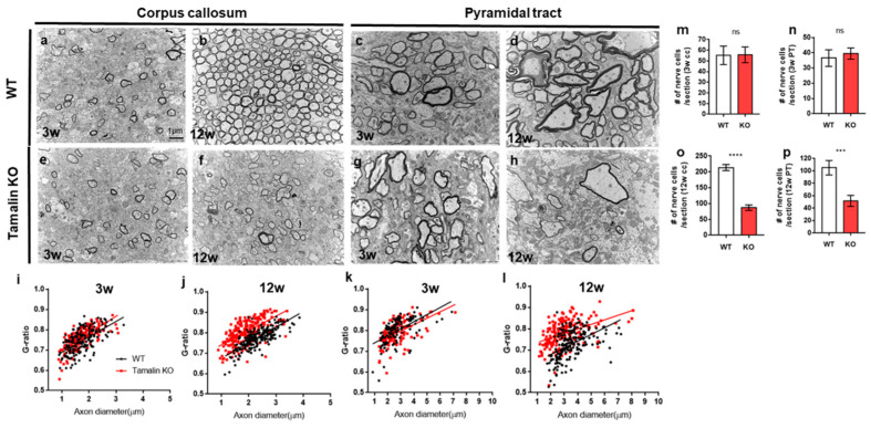
Figure 5 Tamalin KO induces nerve degeneration and hypomyelination in the postembryonic mouse brain. (a–h) TEM images displaying transverse sections of the corpus callosum (a,b,e,f) and pyramidal tract (c,d,g,h) at 3 weeks (a,e,c,g) and 12 weeks (b,f,d,h) in wildtype and tamalin KO mice. (i–l) The g-ratio of myelinated axons in the corpus callosum (i,j) and pyramidal tract (k,l) of wildtype and tamalin KO zebrafish. Unpaired t test was used to compare means from each animal. Each g-ratio was from 100 myelinated axons in eight sections of four mice each (i: p = 0.0535, k: p = 0.0579, j,l: **** p < 0.0001). (m–p) Quantification of the number of nerve cells in the corpus callosum (m,o) and pyramidal tract (n,p) of wildtype and tamalin KO mice (**** p < 0.0001, *** p < 0.001). ns; no significance. Scale bars: (a–h): 1 μm. KO, knockout; TEM, transmission electron microscopy.