
|
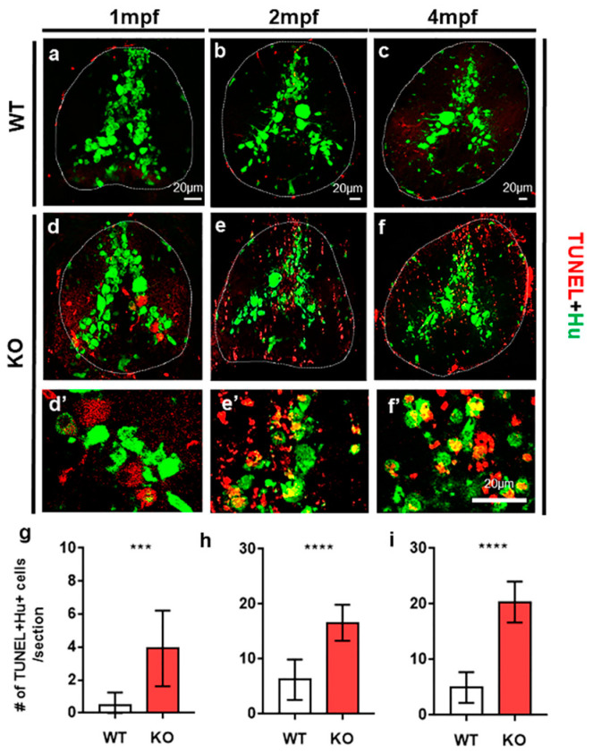
Figure 2 The loss of tamalin function causes neuronal cell death in the spinal cord of postembryonic zebrafish up to adulthood. (a–f) The representative images are transverse sections of the spinal cord of wildtype (a–c) and tamalin KO zebrafish (d–f) labeled with anti-Hu antibody (green) and TUNEL staining (red). (d’–f’) High magnification images of the boxed area in (d–f) show TUNEL+ Hu+ dying neurons. The dorsal side is displayed at the top. (g–i) Quantification of the number of TUNEL+ Hu+ cells in the wildtype or tamalin KO zebrafish at 1 mpf, 2 mpf, and 4 mpf (*** p = 0.0007 **** p < 0.0001, n = 10 sections from five zebrafish). Scale bars: (a–f), 20 µm. KO, knockout; mpf; months post fertilization; and TUNEL, terminal deoxynucleotidyl transferase dUTP nick end labeling.