Fig. 5
Image
Figure Caption
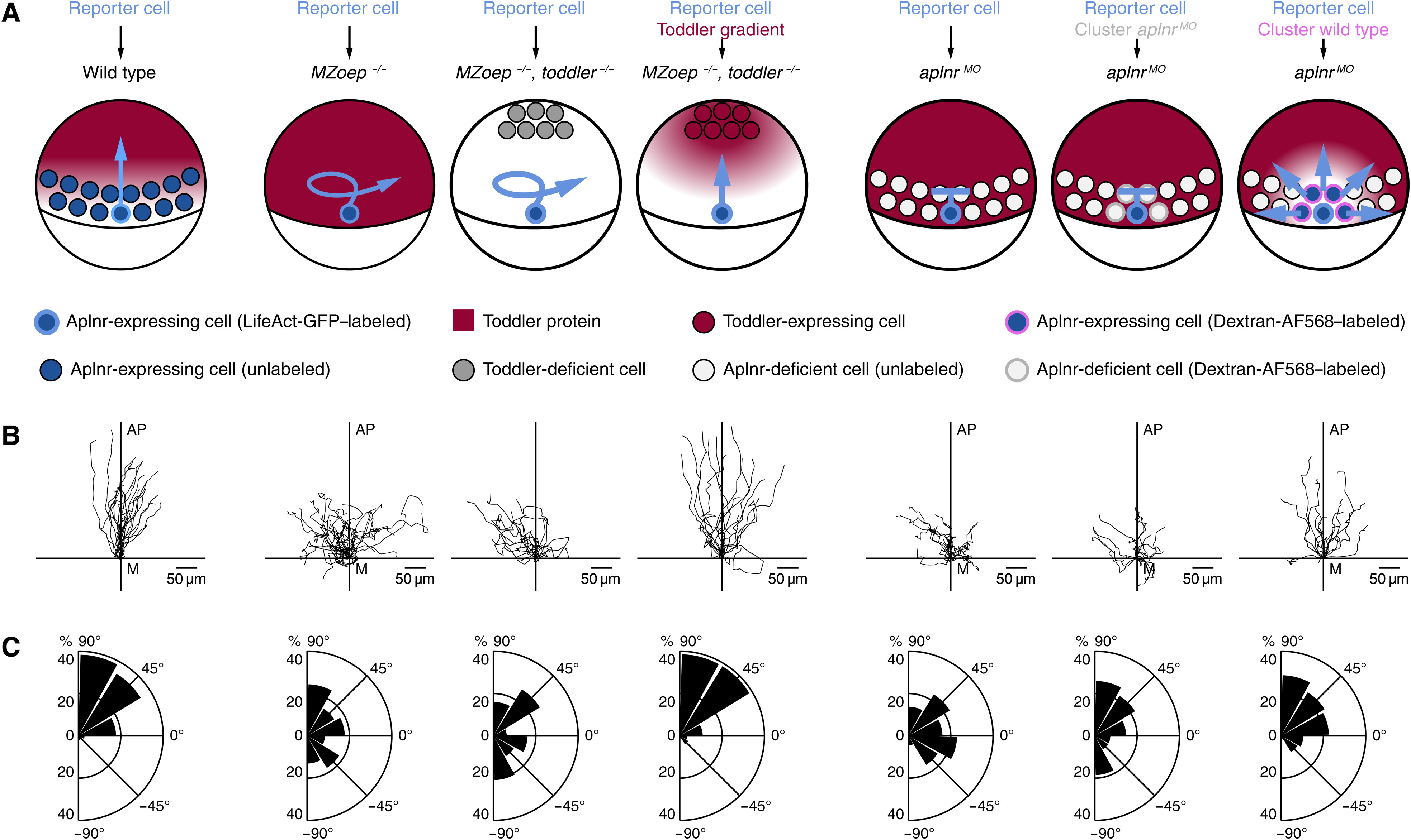
Fig. 5. Aplnr-expressing mesodermal cells are required to establish a Toddler gradient.
Cell transplantation assays to test for the necessity of Aplnr-expressing mesodermal cells as a Toddler sink. Transplanted LifeAct-GFP–labeled wild-type reporter cells were used as a read-out for the presence of a Toddler gradient. (A) Schematic representation of scenarios tested. From left to right: (i) Transplantation of reporter cells into a wild-type host embryo (n = 21). (ii) Transplantation of reporter cells into an MZoep−/− host embryo (n = 22). (iii) Transplantation of reporter cells into an MZoep−/−, toddler−/− double-mutant host embryo. Additional transplantation of Dextran–Alexa Fluor 568–labeled control source cells to the animal pole (n = 16). (iv) Transplantation of reporter cells into an MZoep−/−, toddler−/− double-mutant host embryo. Additional transplantation of Toddler-expressing source cells to the animal pole (n = 15). (v) Transplantation of reporter cells into an aplnr MO embryo (n = 20). (vi) Cotransplantation of one to five reporter cells and a large number of Dextran–Alexa Fluor 568–labeled Aplnr-deficient control cells into aplnrMO host embryo (n = 15). (vii) Cotransplantation of one to five reporter cells and a large number of Aplnr-expressing cells into aplnrMO host embryos (n = 16). (B) Tracks of transplanted reporter cells [order as described in (A)]. Cells were tracked for 90 min after internalization. x axis = margin; y axis = animal-vegetal axis; coordinate origin = start of track. (C) Rose plots showing relative enrichments of orientations of polarity. 90° = animal pole; 0° = ventral/dorsal; −90° = vegetal pole.
Acknowledgments
This image is the copyrighted work of the attributed author or publisher, and
ZFIN has permission only to display this image to its users.
Additional permissions should be obtained from the applicable author or publisher of the image.
Full text @ Sci Adv

