
Fig. 4
- ID
- ZDB-IMAGE-220804-4
- Publication
- Zhang et al., 2022 - Inner nuclear membrane protein TMEM201 maintains endothelial cell migration and angiogenesis by interacting with the LINC complex
- All Figures
- Figures for Zhang et al., 2022
|
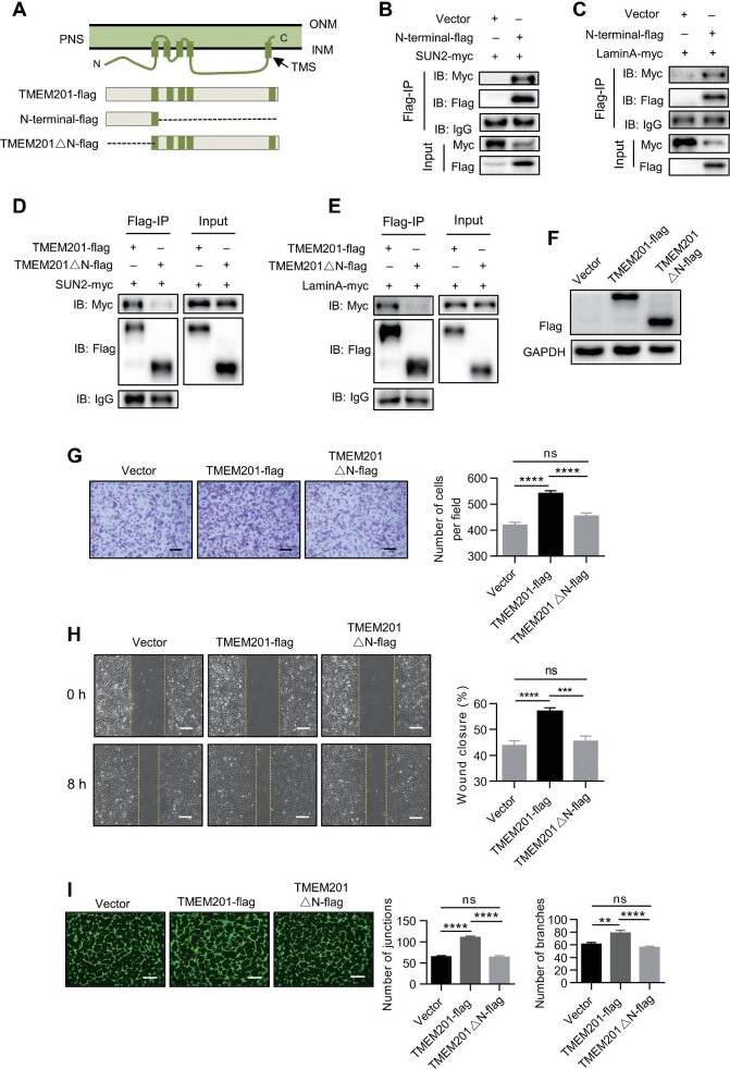
Fig. 4
The N-terminus of TMEM201 interacts with the LINC complex and is required for EC migration. (A) Schematic diagram of Flag-tagged full-length TMEM201 and several truncations for subsequent experiments. (B and C) HEK-293T cells were transfected with vector or TMEM201-N-terminal-flag, together with SUN2-myc (B) or LaminA-myc (C). Cell lysates were immunoprecipitated with anti-Flag and immunoblotted with antibodies against Flag and Myc. (D and E) HEK-293T cells were transfected with full-length TMEM201-flag or TMEM201△N-flag, together with SUN2-myc (D) or LaminA-myc (E). Cell lysates were immunoprecipitated with anti-Flag and immunoblotted with antibodies against Flag and Myc. (F) Western blotting analyses show the relative TMEM201 expression in TMEM201-flag- or TMEM201△N-flag-expressing HUVECs. (G–I) The effects of full-length TMEM201-flag and TMEM201△N-flag in HUVECs were examined using transwell, wound healing, and tube formation assays. (G) Representative images of the transwell assay are shown. Scale bar, 200 μm. The numbers of migrating cells per field were counted. (H) Representative images at 0 and 8 h after scratching are shown. Yellow dotted lines indicate the migration leading edges. The percentage of wound closure was determined at 8 h. Scale bar, 100 μm. (I) Representative fluorescent images and quantitative analyses of the tube formation assay. Tubes were visualized with calcein AM (Green). Scale bar, 200 μm. Data are presented as mean ± SEM (error bars). Statistical significance was determined by one-way ANOVA (**P < 0.01, ***P < 0.001, ****P < 0.0001, and ns = no significance).