
Image
|
Figure Caption
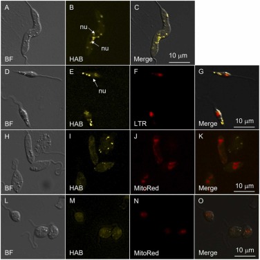
Fig. 3
Fig. 3. Microscopic fluorescence imaging of HAB (5 µM) in L. amazonensis promastigotes in presence of LTR (100 nM) or MitoRed (50 nM). A, D, H, L: bright field imaging; B, E, I, M: HAB labelling; F: LTR labelling; J, N: MitoRed labelling; C, G, K, O: merge. Abbreviations used: nu, nucleus; BF, bright field. The images are representative of three independent experiments.
Acknowledgments
This image is the copyrighted work of the attributed author or publisher, and
ZFIN has permission only to display this image to its users.
Additional permissions should be obtained from the applicable author or publisher of the image.
Full text @ Biomed. Pharmacother.