
|
Fig. 7
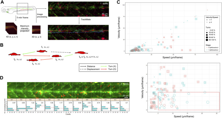
In toto imaging analysis reveals differential cell behaviors and specific spatial distribution of macrophages during neuromast regeneration. (A) The flowchart shows that the trunk region, including neuromast L3-5 was imaged in-toto by light-sheet fluorescence microscopy (LSFM) to monitor macrophage behavior during neuromast regeneration. Larvae from the cross of Tg(mpeg1:mCherry;FoxD3:GFP) and Tg(−8.0cldnb:NTR-hKikGR); Et(HG7L), a wide field of z-stack images focusing around the cloaca (dashed rectangle) was taken every 5 min. We then reduced the order to 3D (x,y,t) with the stacking of z-sections and processed it to have a better resolution by subtracting the background. Finally, we used the plugin “TrackMate” in the Fiji software and manually modified it to acquire the tracking path of every macrophage (shown in the right-bottom corner). (B) We measured the distance (arrows) and displacement (arrows with dashed lines) to calculate speed and Velocity, as shown in (C). Turns were counted among cell movements (indicated by red) to generate the ratio of turns in (C). (C) A scatter plot shows specific speed (the X-axis) and Velocity (the Y-axis) for each macrophage. Other features such as “Speed/Velocity”, “Ratio of turns” and “Stage” are also depicted as shown in the legends on the right to distinguish different cell behaviors better. A region with a speed lower than 25 and a velocity smaller than 10 (dashed rectangle) was magnified, as shown in the lower panel. (D) Histograms divided by sections (width of 100 μm) represent counts (the X-axis) of the different numbers of recruited macrophages (the Y-axis) during neuromast regeneration. In each panel, the mean of the “number of recruited macrophages” is shown in the right-upper graph and presented with dashed lines in red.