
|
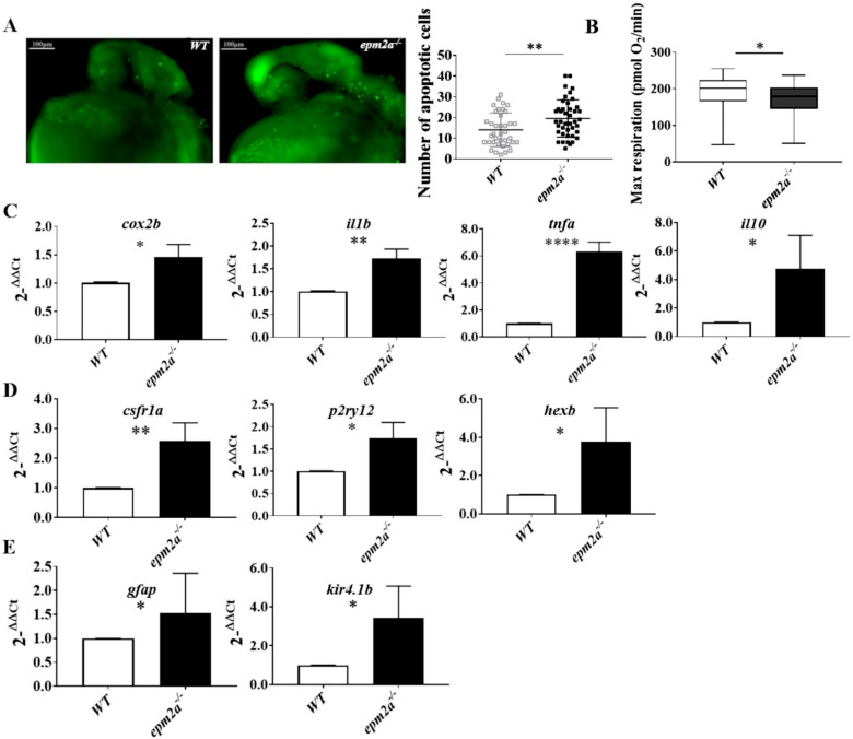
Fig. 6
Neuroinflammation, apoptotic death, and mitochondrial dysfunction in epm2a−/− zebrafish larvae. (A) Detection of apoptotic cells by acridine orange staining in controls and epm2a−/− mutant embryos at 24 hpf (lateral views) alongside a quantitative analysis of apoptotic cells in WT (n = 40) and epm2a−/− (n = 44) fish. ** p ≤ 0.01, calculated by the Mann-Whitney test. (B) Mitochondrial respiratory analysis of controls (n = 47) and epm2a−/− mutant larvae (n = 48) at 120 hpf. * p ≤ 0.05, calculated by the Mann-Whitney test. (C) qRT–PCR analysis of inflammatory and anti-inflammatory cytokines. (D) qRT–PCR analysis of microglial genes. (E) qRT–PCR analysis of astroglial genes. The mRNA expression levels had been normalized to expression of β-actin. Three independent RNA samples (each obtained from about 30–40 larvae) from controls and epm2a−/− mutant larvae at 120 hpf were analyzed. * p ≤ 0.05, ** p ≤ 0.01, **** p ≤ 0.0001, calculated by Student’s t-test. The values are expressed as mean ± SD.