
Image
|
Figure Caption
Fig. 8
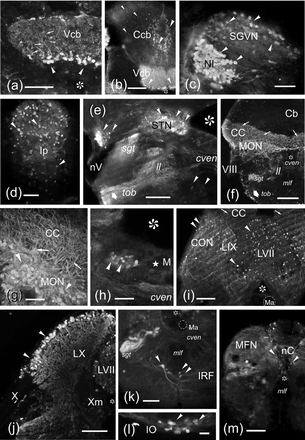
(a–m) Photomicrographs of transverse sections of the zebrafish rhombencephalon showing Nrgn-like-ir cell bodies (arrowheads) and processes (arrows). Medial is to the right except in (d). (a,b) Nrgn-like-ir in Purkinje cells in the medial division of the valvula (a, b) and corpus (b) of the cerebellum. Only in the cerebellar valvula (Vcb) the apical dendrites of these cells (arrows) can be well observed. (c) Detail of the NI and the secondary gustatory-visceral nucleus (SGVN) showing intensely and lightly labeled immunoreactive cell bodies, respectively. (d) Detail of the interpeduncular nucleus (Ip). (e) Nrgn-like-ir neurons in the sensory nucleus of the trigeminal nerve (STN) and dorsal to the sensory root entrance of the trigeminal nerve (nV). (f) General view of a cryostat section at the level of the octaval nerve (VIII) entrance. Note the high amount of Nrgn-like-ir dendrites in the CC and the Nrgn-like-ir in two fiber bundles, the secondary gustatory tract (sgt) and the bundle at the ventrolateral margin of the tegmentum (thick arrow). (g) Vibratome cross section showing a detail of the Nrgn-like-ir cells of the MON. (h) Vibratome section showing a discrete group of Nrgn-like-ir cells close to the Mauthner cell. (i) Nrgn-like-ir cells (arrowheads) in the caudal octavolateral region and the viscerosensory lobes (IX, VII). Note the Nrgn-like-ir dendrites of the CON (arrows) entering the CC. (j) Detail of the vagal lobe showing Nrgn-like-ir cell bodies mostly at its periphery. (k) Vibratome cross section at the level of the inferior reticular formation showing immunoreactivity in a large reticular cell and a couple of small cells. Note also the sgt intensely labeled. (l) Detail of Nrgn-like-ir cell bodies in the IO. (m) Section at the level of the commissural nucleus of Cajal (nC). Asterisk, ventricle. For abbreviations, see the list. Scale bars: 500 μm (f), 200 μm (k, m), 100 μm (a,b,e, i,j), 50 μm (c,d, g,h),and20 μm (l)
Figure Data
Acknowledgments
This image is the copyrighted work of the attributed author or publisher, and
ZFIN has permission only to display this image to its users.
Additional permissions should be obtained from the applicable author or publisher of the image.
Full text @ J. Comp. Neurol.