
- Title
-
Distribution of Neurogranin-like immunoreactivity in the brain and sensory organs of the adult Zebrafish
- Authors
- Alba-González, A., Folgueira, M., Castro, A., Anadón, R., Yáñez, J.
- Source
- Full text @ J. Comp. Neurol.
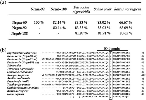
(a) Results of NCBI Blastp showing amino acid identity (%) of full-length sequences of neurogranin from rat (R. norvegicus), spotted green pufferfish (T. nigroviridis), Atlantic salmon (S. salar), and zebrafish (D. rerio) variants a (derived from nrgna > Nrgna-60 and Nrgna-92) and variant b (nrgnb > Nrgnb-188). (b) Alignment of neurogranin sequences containing IQ domain between organisms. Right column indicates the position of the last amino acid shown in the selected portion of the whole sequence. Twenty-five amino acids were fully conserved (*), six amino acids were strongly similar (:) and only two amino acids were weakly similar (.). IQ domain is highly conserved in all sequences [16 (*), 2 (:), and 1 (.)]. Serine (S) located in position 36 (outlined box) in basal actinopterigyans (E. calabaricus), reptiles, birds, and mammals, is substituted by glycine (G) in teleosts and X. tropicalis, and by asparagine (N) in sarcopterygii (L. chalumnae)
|
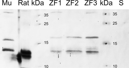
Western blot results for the different protein homogenates employed from zebrafish (ZF and S), Wistar rat (Rat) and gray mullet (Mu). Two zebrafish brain extracts (ZF1 and ZF3) show three Nrgn-ir bands of about 13, 20 and 37 kDa. Note the absence of the 37-kDa band in the zebrafish brain extract lacking the telencephalic lobes (ZF2). In zebrafish spleen homogenate (S), no Nrgn-ir bands were observed. Immunoblotting with anti-Nrgn-antiserum was also analyzed in parallel in brain extracts of Wistar rat (R. norvegicus) and gray mullet (Mu; C. labrosus)
|
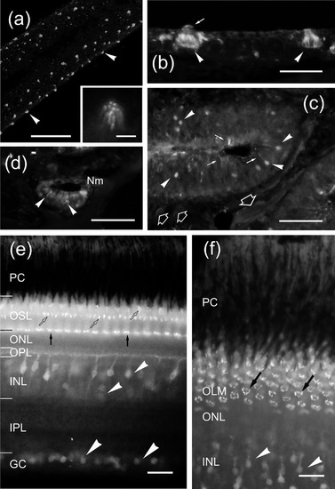
(a–d) Nrgn-like distribution in the surface of a barbel (a), skin (b), the olfactory organ (c) and the retina (e, f). (a) Nrgn-like immunoreactivity in the apical region of sensory cells in taste buds (arrowheads); detailed in the inset. (b) Transverse section of two Nrgn-like immunoreactive taste buds (arrowheads) at the dorsal surface of the head. Arrow points to apical microvilli. (c) Transverse section of the olfactory rosette showing Nrgn-like-ir in cell bodies (arrowheads) and apical dendrites (arrows) of olfactory receptor cells. Note the very low immunoreactivity in the olfactory axon bundles (open arrows). (d) Detail of a supraorbital canal neuromast showing weak Nrgn-like immunoreaction in hair cells (arrowheads). (e) Vertical section of the retina showing Nrgn-like-ir cell bodies (arrowhead), puncta (black arrows) and comma-like structures (white arrows). (f) Oblique section through the retina showing Nrgn-like-ir cell bodies in the INL (arrowheads) and details of the Nrgn-like-ir puncta arranged in a ring pattern around the base of the inner segment of photoreceptors (black arrows). For abbreviations, see the list. Scale bars: 100 μm (a–c), 50 μm (b,d), 25 μm (e–f), and 10 μm (inset in a)
EXPRESSION / LABELING:
|
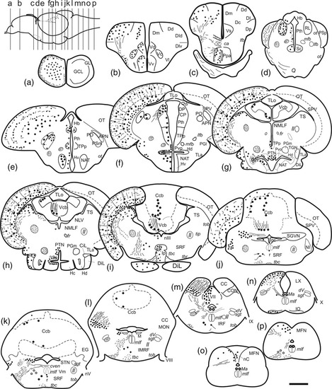
(a–p) Schematic drawings of transverse sections of the zebrafish brain from rostral (a) to caudal (p) showing on the left the Nrgn-like-ir cell bodies (big dots), neuronal processes and tracts (thin lines and small dots), while on the right the annotations of different nuclei and tracts. The levels of the sections are indicated by lines in the lateral diagram of the brain on the left. For abbreviations, see the list. Scale bar for sections: 200 μm (a–p)
EXPRESSION / LABELING:
|
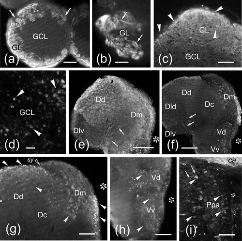
(a–i) Photomicrographs of transverse sections of the olfactory bulb (a–d), telencephalic lobes (e–h) and preoptic area (i) of the zebrafish brain showing Nrgn-like-ir cell bodies (arrowheads) and fibers (arrows). Medial is to the right. (a) General view of a vibratome section of the olfactory bulb showing Nrgn-like-ir fibers in the glomerular layer. (b) Detail of an olfactory glomerulus showing Nrgn-like-ir fibers. (c) Detail of a cryostat section showing Nrgn-like-ir cell bodies in the glomerular layer. (d) Detail of Nrgn-like-ir granule cells. (e) General view of a telencephalic lobe at the rostral level. (f) Vibratome section of a telencephalic lobe at precommissural level. (g) Detail of a vibratome section showing Nrgn-like-ir cell bodies. Note the absence of labeled cell bodies in the Dlv and Dc. (h) Vibratome section through the subpallium showing positive cell bodies in Vd and Vv. (i) Detail of positive cell bodies in the anterior parvocellular preoptic region (Ppa). Asterisk, ventricle. For abbreviations, see the list. Scale bars: 200 μm (e–f), 100 μm (a, g–h), 50 μm (b,c, i), and 20 μm (d)
EXPRESSION / LABELING:
|
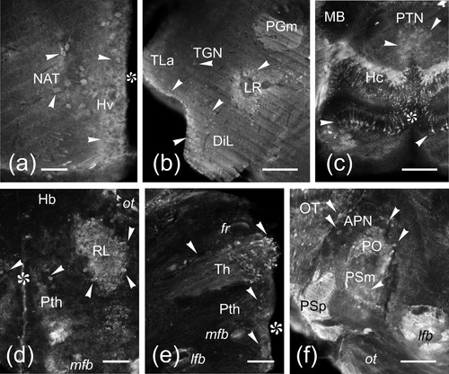
(a–f) Vibratome (a–c, e–f) and cryostat (d) cross sections of the zebrafish brain through the hypothalamus (a,b) and diencephalon (c–f), showing Nrgn-like-ir labeled cell bodies (arrowheads) and processes (arrows). Medial is to the right. (a) Section through the rostral hypothalamic region showing lightly labeled Nrgn-like-ir cell bodies in the NAT. (b) Detail of the lateral inferior lobe showing immunopositive cells in the torus lateralis (TLa), diffuse nucleus (DiL) and around the lateral recess (LR). (c) Detail of Nrgn-like-ir immunoreaction in the posterior lobe. (d) Section showing immunoreactive cell bodies in the nucleus rostrolateralis (RL). (e) Section through the thalamus. (f) Detail of the pretectal area. Asterisk, ventricle. For abbreviations, see the list. Scale bars: 100 μm (b,c, e–f), 50 μm (a,d)
EXPRESSION / LABELING:
|
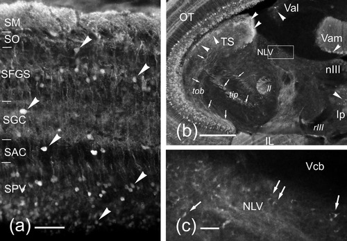
(a–c) Photomicrographs of transverse sections through the mesencephalon of the zebrafish showing Nrgn-like-ir immunoreaction in cell bodies (arrowheads) and processes (arrows). (a) Cross section through the OT showing immunoreactive cell bodies (arrowheads). Note the SM is crowded with immunoreactive dendritic branches. (b) Transverse vibratome section through the mesencephalic tegmentum. The midline is to the right. (c) Detail of Nrgn-like-ir axonal endings (arrows) in the NLV. For abbreviations, see the list. Scale bars: 200 μm (b), 50 μm (a), 20 μm (c)
EXPRESSION / LABELING:
|
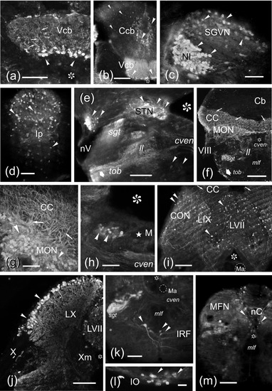
(a–m) Photomicrographs of transverse sections of the zebrafish rhombencephalon showing Nrgn-like-ir cell bodies (arrowheads) and processes (arrows). Medial is to the right except in (d). (a,b) Nrgn-like-ir in Purkinje cells in the medial division of the valvula (a, b) and corpus (b) of the cerebellum. Only in the cerebellar valvula (Vcb) the apical dendrites of these cells (arrows) can be well observed. (c) Detail of the NI and the secondary gustatory-visceral nucleus (SGVN) showing intensely and lightly labeled immunoreactive cell bodies, respectively. (d) Detail of the interpeduncular nucleus (Ip). (e) Nrgn-like-ir neurons in the sensory nucleus of the trigeminal nerve (STN) and dorsal to the sensory root entrance of the trigeminal nerve (nV). (f) General view of a cryostat section at the level of the octaval nerve (VIII) entrance. Note the high amount of Nrgn-like-ir dendrites in the CC and the Nrgn-like-ir in two fiber bundles, the secondary gustatory tract (sgt) and the bundle at the ventrolateral margin of the tegmentum (thick arrow). (g) Vibratome cross section showing a detail of the Nrgn-like-ir cells of the MON. (h) Vibratome section showing a discrete group of Nrgn-like-ir cells close to the Mauthner cell. (i) Nrgn-like-ir cells (arrowheads) in the caudal octavolateral region and the viscerosensory lobes (IX, VII). Note the Nrgn-like-ir dendrites of the CON (arrows) entering the CC. (j) Detail of the vagal lobe showing Nrgn-like-ir cell bodies mostly at its periphery. (k) Vibratome cross section at the level of the inferior reticular formation showing immunoreactivity in a large reticular cell and a couple of small cells. Note also the sgt intensely labeled. (l) Detail of Nrgn-like-ir cell bodies in the IO. (m) Section at the level of the commissural nucleus of Cajal (nC). Asterisk, ventricle. For abbreviations, see the list. Scale bars: 500 μm (f), 200 μm (k, m), 100 μm (a,b,e, i,j), 50 μm (c,d, g,h),and20 μm (l)
EXPRESSION / LABELING:
|