
Image
|
Figure Caption
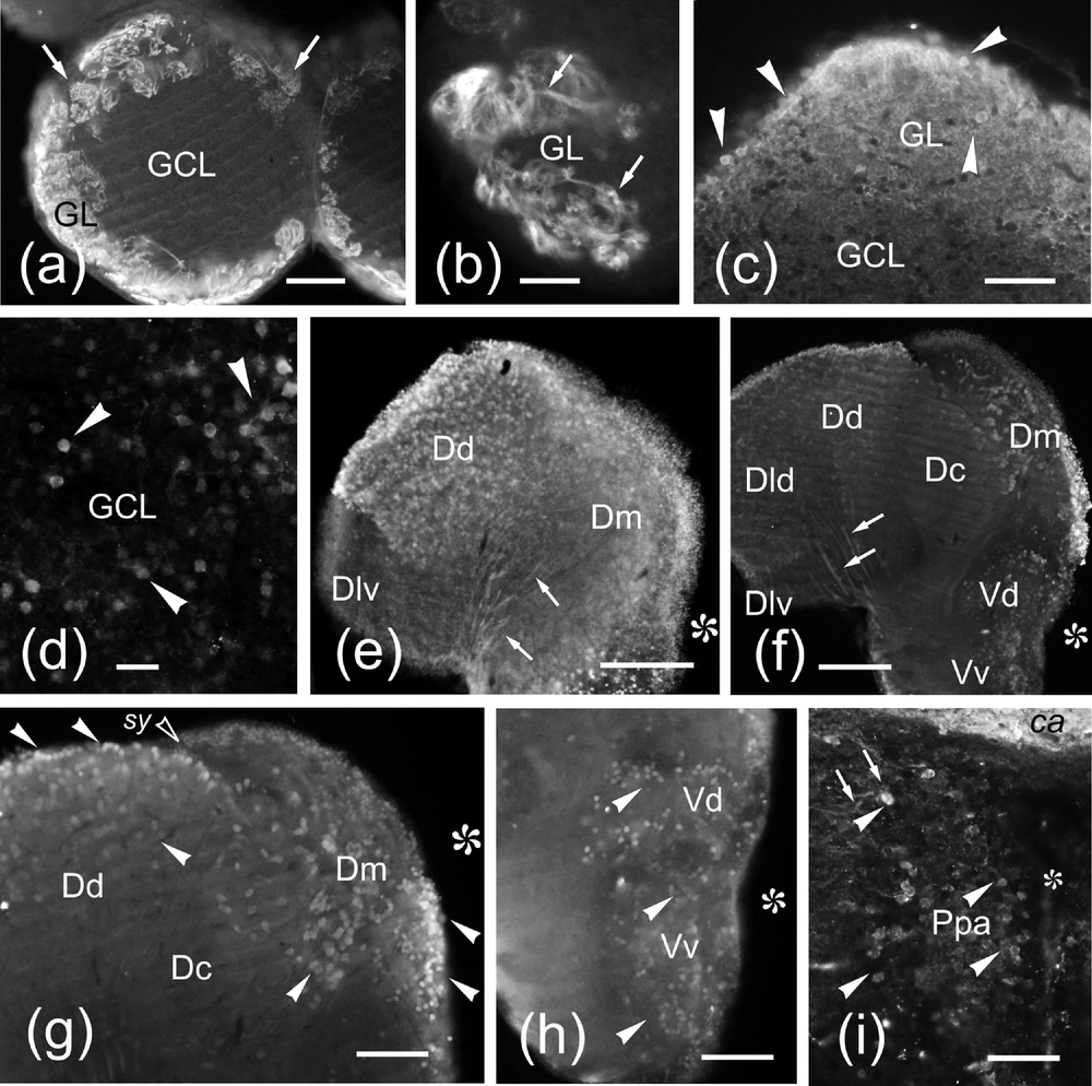
Fig. 5
(a–i) Photomicrographs of transverse sections of the olfactory bulb (a–d), telencephalic lobes (e–h) and preoptic area (i) of the zebrafish brain showing Nrgn-like-ir cell bodies (arrowheads) and fibers (arrows). Medial is to the right. (a) General view of a vibratome section of the olfactory bulb showing Nrgn-like-ir fibers in the glomerular layer. (b) Detail of an olfactory glomerulus showing Nrgn-like-ir fibers. (c) Detail of a cryostat section showing Nrgn-like-ir cell bodies in the glomerular layer. (d) Detail of Nrgn-like-ir granule cells. (e) General view of a telencephalic lobe at the rostral level. (f) Vibratome section of a telencephalic lobe at precommissural level. (g) Detail of a vibratome section showing Nrgn-like-ir cell bodies. Note the absence of labeled cell bodies in the Dlv and Dc. (h) Vibratome section through the subpallium showing positive cell bodies in Vd and Vv. (i) Detail of positive cell bodies in the anterior parvocellular preoptic region (Ppa). Asterisk, ventricle. For abbreviations, see the list. Scale bars: 200 μm (e–f), 100 μm (a, g–h), 50 μm (b,c, i), and 20 μm (d)
Figure Data
Acknowledgments
This image is the copyrighted work of the attributed author or publisher, and
ZFIN has permission only to display this image to its users.
Additional permissions should be obtained from the applicable author or publisher of the image.
Full text @ J. Comp. Neurol.