
Image
|
Figure Caption
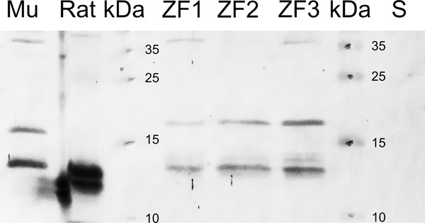
Fig. 2
Western blot results for the different protein homogenates employed from zebrafish (ZF and S), Wistar rat (Rat) and gray mullet (Mu). Two zebrafish brain extracts (ZF1 and ZF3) show three Nrgn-ir bands of about 13, 20 and 37 kDa. Note the absence of the 37-kDa band in the zebrafish brain extract lacking the telencephalic lobes (ZF2). In zebrafish spleen homogenate (S), no Nrgn-ir bands were observed. Immunoblotting with anti-Nrgn-antiserum was also analyzed in parallel in brain extracts of Wistar rat (R. norvegicus) and gray mullet (Mu; C. labrosus)
Figure Data
Acknowledgments
This image is the copyrighted work of the attributed author or publisher, and
ZFIN has permission only to display this image to its users.
Additional permissions should be obtained from the applicable author or publisher of the image.
Full text @ J. Comp. Neurol.