IMAGE

Figure 4.

ID
ZDB-IMAGE-220414-17
Source
Figures for Tallafuss et al., 2022
Image
Figure Caption

Figure 4.

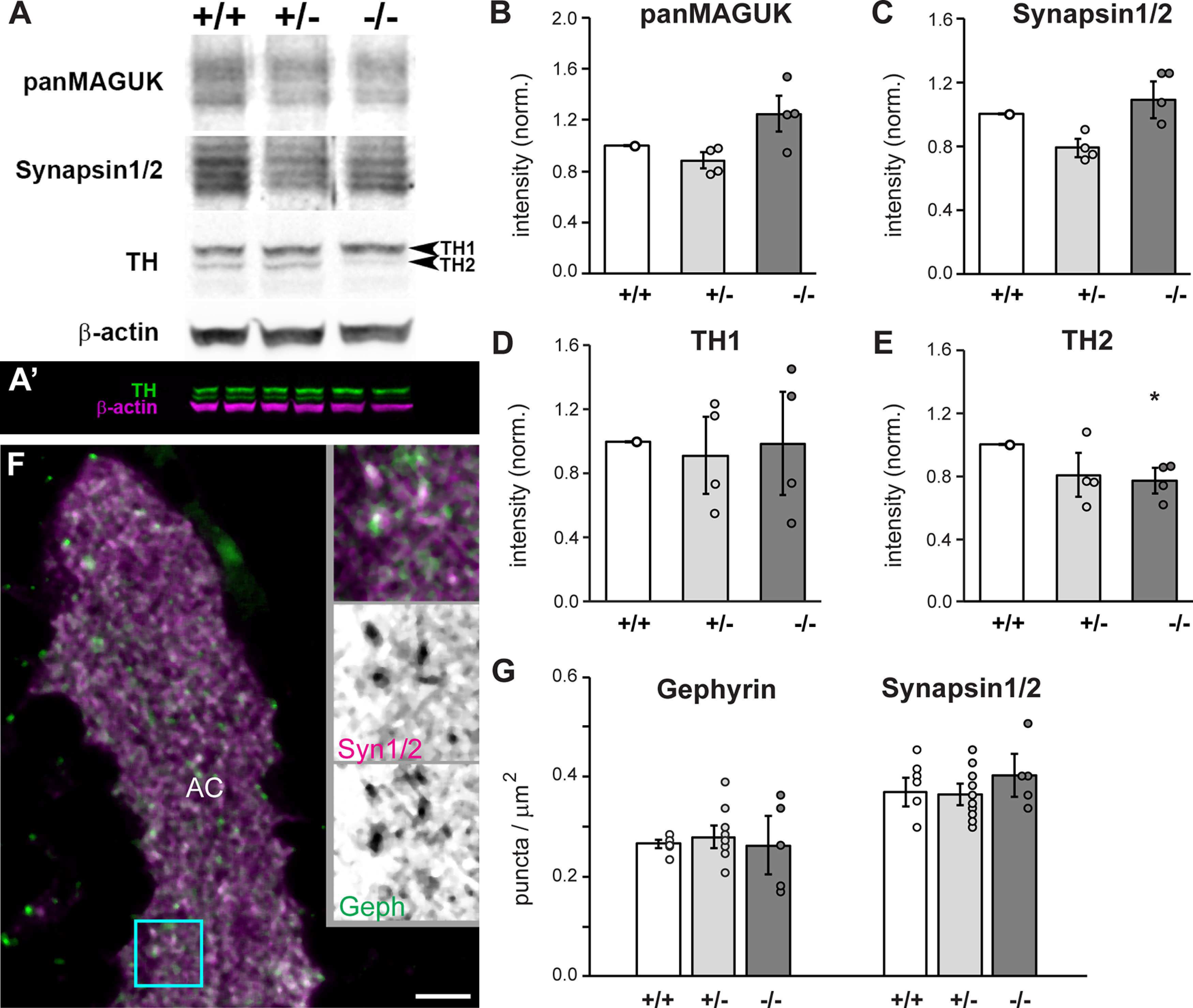
Egr1 is necessary for TH2 expression. A, Western blotting of larval head homogenates at 14 dpf reveals a reduction in expression of TH2 in egr1 mutants. A’, Two color labeling of a Western blotting to highlight in-lane normalization to β-actin and technical replicates. B–E, Western blot intensities in A, A’ were first normalized to in-lane β-actin, and then to wt average intensity. N = 4 experiments, n = 2 technical replicates, *p < 0.05, HSD. F, Immunolabeling of synapses with antibodies to Synapsin 1 and 2 (Syn1/2, magenta) and Gephyrin (Geph, green) in the anterior commissure (AC), lateral view. Insets are magnifications of the cyan box. Scale bar: 5 μm. G, Quantification of synaptic puncta revealed no changes in synapse density in the medial and lateral AC. n ≥ 5 brains per genotype. wt (+/+, white), het (+/−, light gray), mut (−/−, dark gray). Error bars are SEM.

Acknowledgments
This image is the copyrighted work of the attributed author or publisher, and ZFIN has permission only to display this image to its users. Additional permissions should be obtained from the applicable author or publisher of the image. Full text @ eNeuro