IMAGE

Figure 1.

ID
ZDB-IMAGE-220414-14
Source
Figures for Tallafuss et al., 2022
Image
Figure Caption

Figure 1.

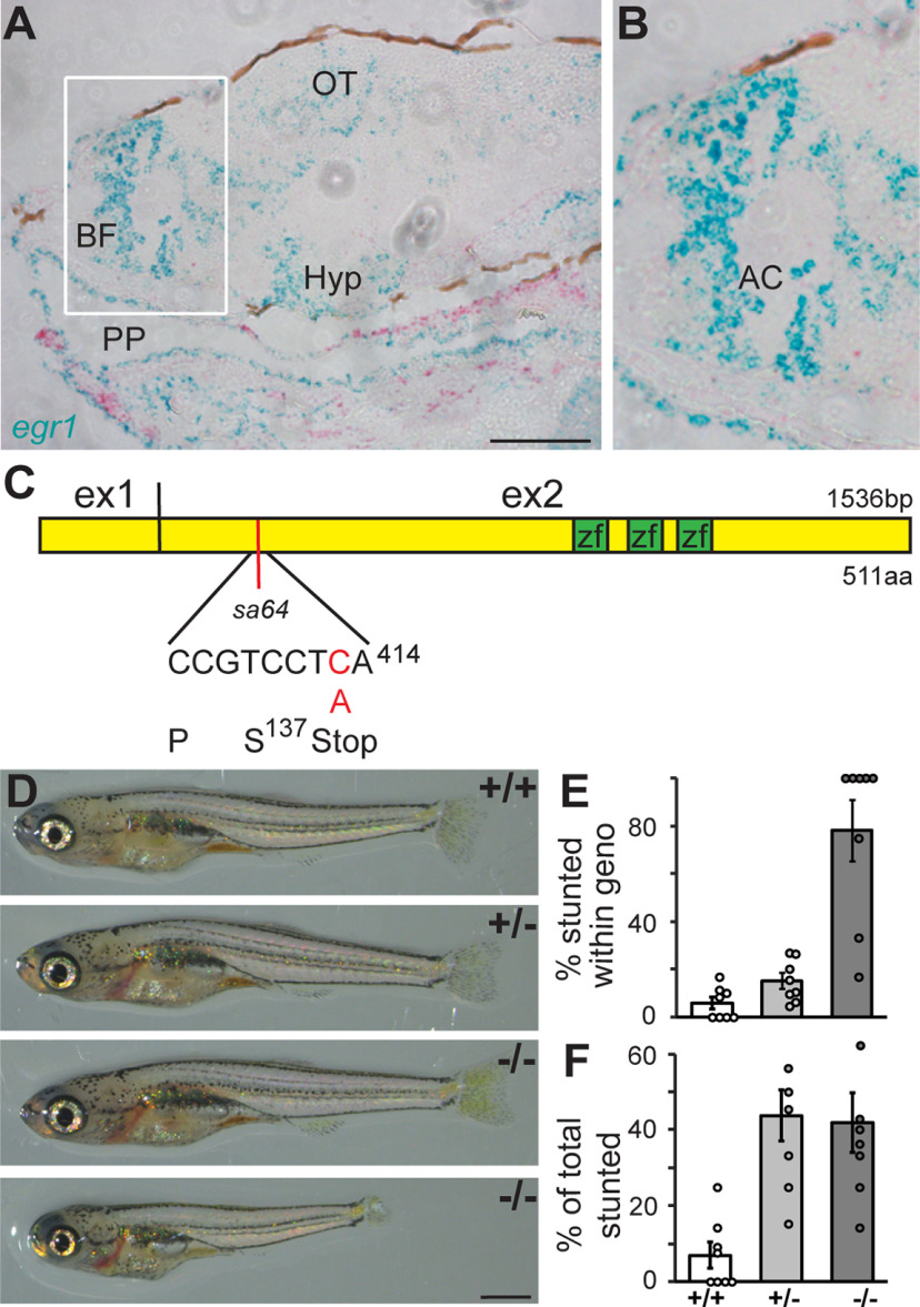
egr1 is expressed in the forebrain. A, ISH reveals egr1 expression (blue) in the basal forebrain (BF), optic tectum (OT), hypothalamus (Hyp), and epithelial cells of the pharyngeal pouch (PP) at 6 dpf. myogenin d was used as a control probe (red). Anterior to the left. Scale bar: 100 μm. B, Magnification of the BF (white box in A) showing expression surrounding the anterior commissure (AC). C, Exon organization of the egr1 mRNA, location of the zinc finger domains (zf), and location of the sa64 mutation (red bar). D, egr1sa64 mutants (−/−) develop a normal body plan with functional swim bladder, but with size deficits (bottom). E, Close to 80% of mutant larvae are stunted (dark gray). F, More than 50% of stunted larvae are also wt (+/+, white) and het animals (+/−, light gray). N = 8 clutches, n = 248 animals.

Acknowledgments
This image is the copyrighted work of the attributed author or publisher, and ZFIN has permission only to display this image to its users. Additional permissions should be obtained from the applicable author or publisher of the image. Full text @ eNeuro