IMAGE

Fig 2

ID
ZDB-IMAGE-220319-51
Source
Figures for Liu et al., 2022
Image
Figure Caption

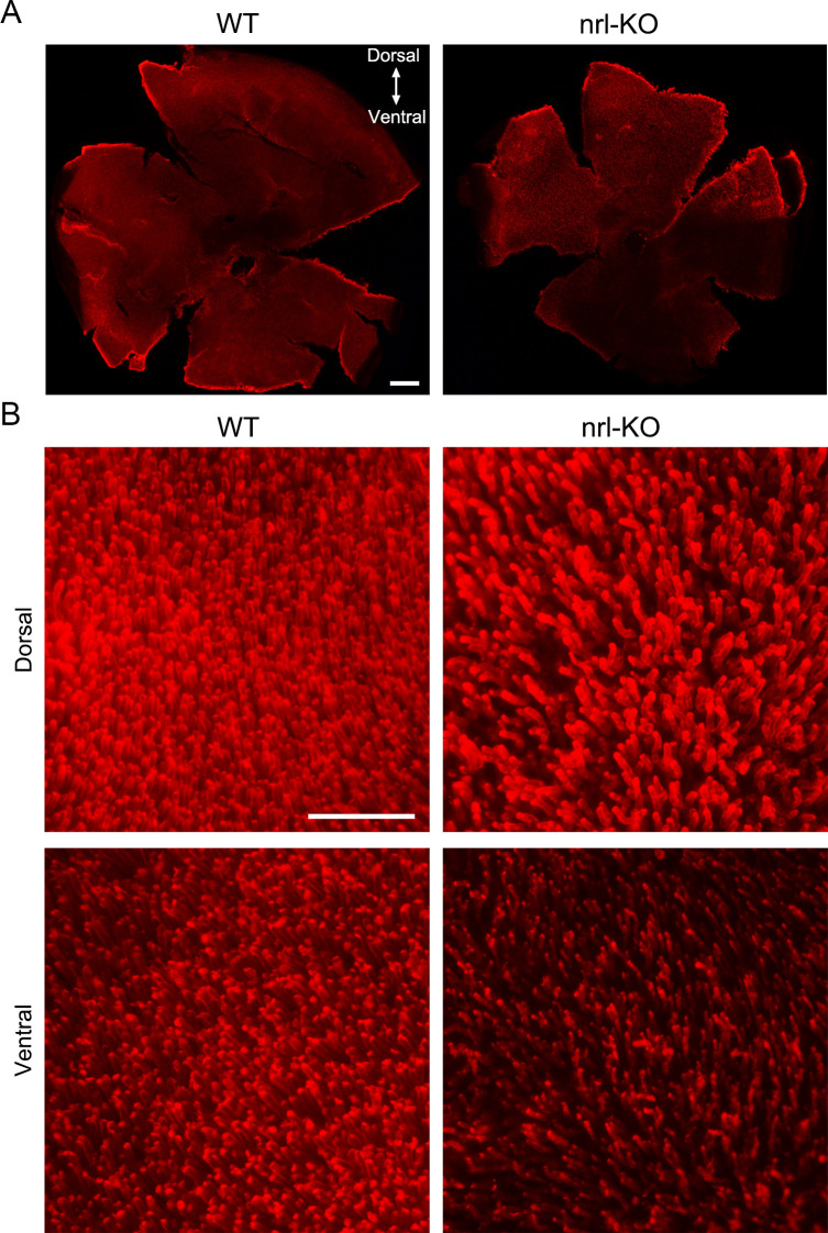
Fig 2 Reduced density of the rod outer segments in the <italic toggle='yes'>nrl</italic>-KO zebrafish.

(A) The rod outer segments were visualized via immunostaining on the flattened whole-mount retinas from WT and nrl-KO zebrafish at 2 mpf using the anti-Rho antibody. The overall views of WT and nrl-KO retinas are shown in the low-magnification images. Scale bar: 200 μm. (B) High-magnification images from the dorsal and ventral retinal regions are shown. The density of rods is reduced in the nrl knockout retinas, especially in the ventral retinal region. Scale bar: 50 μm.

Figure Data
Acknowledgments
This image is the copyrighted work of the attributed author or publisher, and ZFIN has permission only to display this image to its users. Additional permissions should be obtained from the applicable author or publisher of the image. Full text @ PLoS Genet.